. 2010 Jun 15;5(6):e11131.
doi: 10.1371/journal.pone.0011131.

Assessment of the potential role of muscle spindle mechanoreceptor afferents in chronic muscle pain in the rat masseter muscle

Affiliations

Assessment of the potential role of muscle spindle mechanoreceptor afferents in chronic muscle pain in the rat masseter muscle

James P Lund et al. PLoS One. .

Abstract

Background: The phenotype of large diameter sensory afferent neurons changes in several models of neuropathic pain. We asked if similar changes also occur in "functional" pain syndromes.

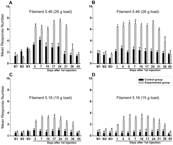
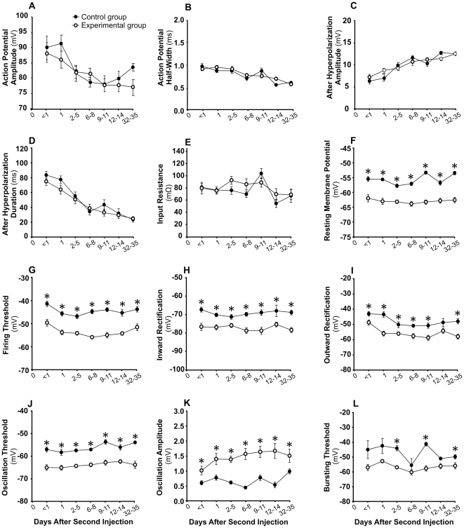
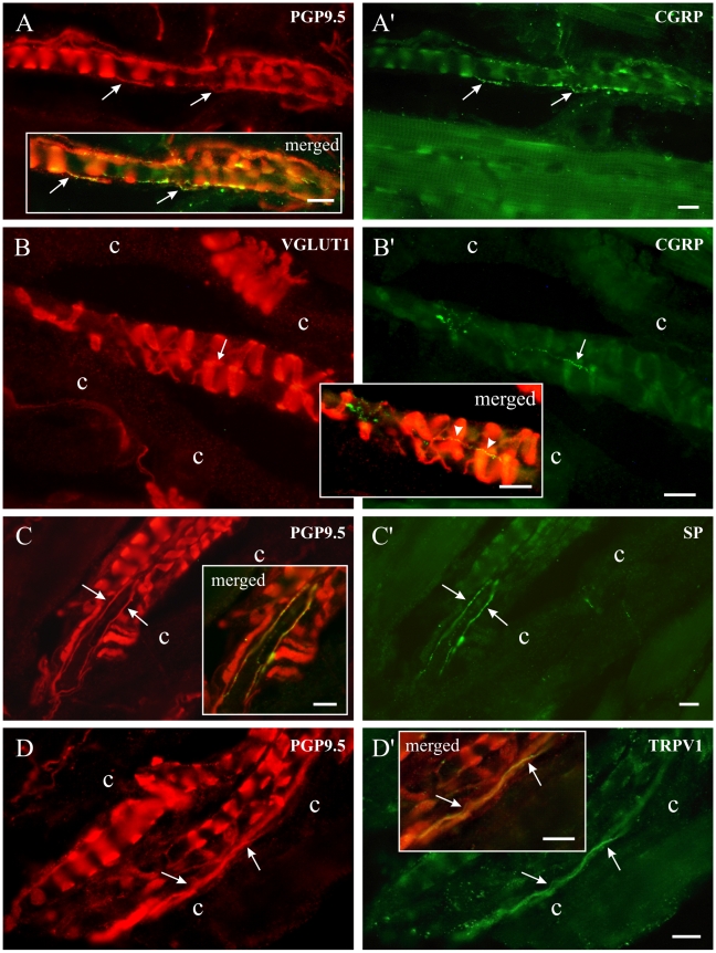
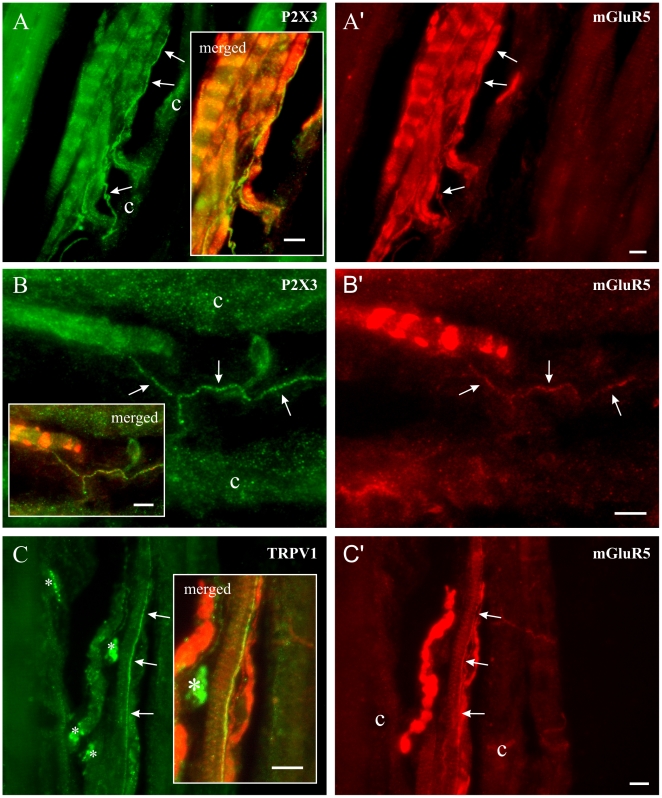
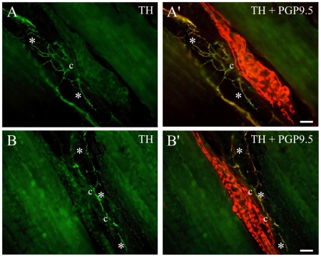
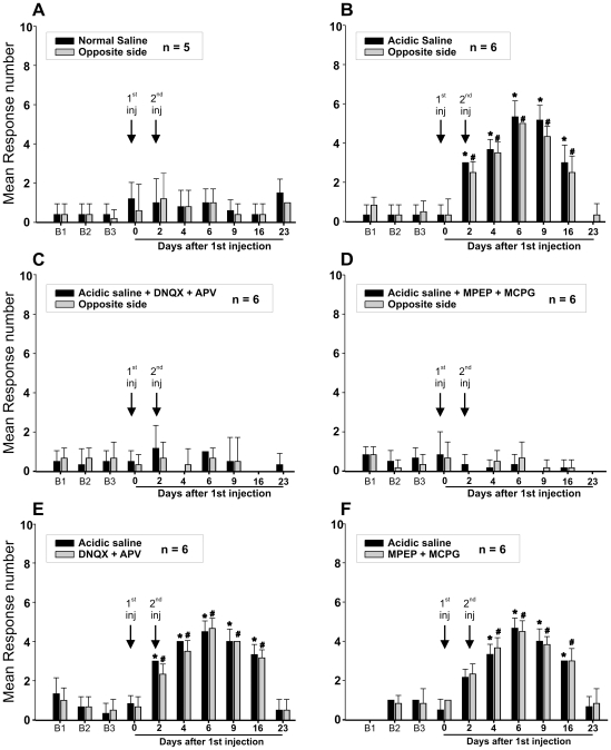
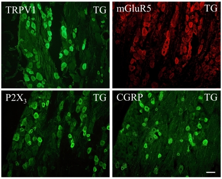
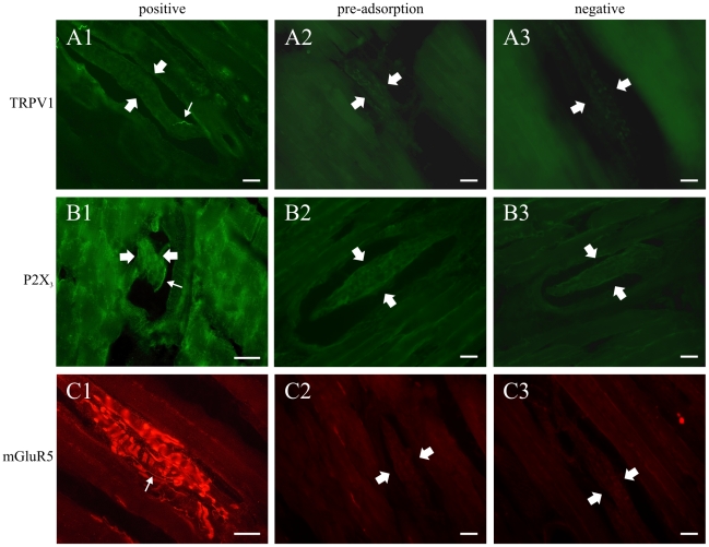
Methodology/principal findings: Acidic saline (AS, pH 4.0) injections into the masseter muscle were used to induce persistent myalgia. Controls received saline at pH 7.2. Nocifensive responses of Experimental rats to applications of Von Frey Filaments to the masseters were above control levels 1-38 days post-injection. This effect was bilateral. Expression of c-Fos in the Trigeminal Mesencephalic Nucleus (NVmes), which contains the somata of masseter muscle spindle afferents (MSA), was above baseline levels 1 and 4 days after AS. The resting membrane potentials of neurons exposed to AS (n = 167) were hyperpolarized when compared to their control counterparts (n = 141), as were their thresholds for firing, high frequency membrane oscillations (HFMO), bursting, inward and outward rectification. The amplitude of HFMO was increased and spontaneous ectopic firing occurred in 10% of acid-exposed neurons, but never in Controls. These changes appeared within the same time frame as the observed nocifensive behaviour. Ectopic action potentials can travel centrally, but also antidromically to the peripheral terminals of MSA where they could cause neurotransmitter release and activation of adjacent fibre terminals. Using immunohistochemistry, we confirmed that annulospiral endings of masseter MSA express the glutamate vesicular transporter VGLUT1, indicating that they can release glutamate. Many capsules also contained fine fibers that were labelled by markers associated with nociceptors (calcitonin gene-related peptide, Substance P, P2X3 receptors and TRPV1 receptors) and that expressed the metabotropic glutamate receptor, mGluR5. Antagonists of glutamatergic receptors given together with the 2(nd) injection of AS prevented the hypersensitivity observed bilaterally but were ineffective if given contralaterally.

Conclusions/significance: Low pH leads to changes in several electrical properties of MSA, including initiation of ectopic action potentials which could propagate centrally but could also invade the peripheral endings causing glutamate release and activation of nearby nociceptors within the spindle capsule. This peripheral drive could contribute both to the transition to, and maintenance of, persistent muscle pain as seen in some "functional" pain syndromes.


Conflict of interest statement

Competing Interests: The authors have declared that no competing interests exist.

Figures

Figure 1
Figure 1. Acidic saline injections increase nocifensive behaviour.
Animals received two injections of either normal saline (Control group, black bars) or acidic saline (Experimental group, gray bars) into both masseter muscles. The injections were made 2 days apart. Pressure was applied to the centre of the masseter muscle with von Frey filaments, and flinching and/or head withdrawal was scored as a positive response. Each muscle was tested 10 times with each of the filaments, and data from left and right were averaged. Means and standard errors are shown for three days prior to injection (B1, B2 and B3), and for 45 days after the first of the two injections. A, C: postinjection testing began 2 days after the second injection (n = 6). B, D: testing of these animals (n = 6) began 1 day after the first injection. *, p<0.05.
Figure 2
Figure 2. Normal and acidic saline injections increase the number of c-Fos expressing cells in NVmes, but only with acidic saline does the level of expression remain elevated.
Graph showing the mean number ± S.E. of c-Fos stained NVmes cells in Unoperated Controls (open diamonds), and Experimental (open squares) and Control group (filled circles) at 4, 24 and 96 hours after injection. Asterisks above points: significance compared to the Control group. Asterisks below points: significance compared to Unoperated Controls. *, p<0.05; **, p<0.01.
Figure 3
Figure 3. Acidic saline injections produce long lasting changes in many of the electrophysiological properties of NVmes cells innervating masseter muscle spindles.
Means ± S.E. of twelve electrical properties are shown for Control (filled circle) and Experimental (open circle) neurons recorded at 7 time periods after the second intramuscular injection. *, p<0.05 for simple contrasts. In panels F, G and J, all ps<0.01.
Figure 4
Figure 4
A: At equivalent membrane potentials, the subthreshold membrane oscillations of NVmes cells exposed to acidic saline are larger and the number of cells firing repetitively is greater. Examples of recordings made 14 days after the second injection in Control (left) and Experimental neurons showing the difference in oscillation amplitude at similar membrane potentials. The amount of current injected is given on the right of each trace. Calibration bars in the Control panel also apply to the Experimental panel. B: Examples of the four types of firing patterns induced by current injections. The inset illustrates a section of the “Bursting” trace at higher magnification to show that the bursts coincided with period of high amplitude oscillations. Calibration bars in top left panel apply to all panels in B. RMP: resting membrane potential.
Figure 5
Figure 5. Masseter muscle spindles contain small-calibre afferents that express nociceptor markers.
Left and right photomicrographs have identical frames with different sets of fluorescence filters. In each case, portions of the photomicrographs were digitally merged in boxed areas. A: Nerve fibres immunoreactive for PGP9.5 and containing CGRP (A'). B: A small green CGRP-positive fibre (B', thin arrow) runs across three VGLUT1-positive loops of an annulospiral ending. None of the VGLUT1-positive fibres in B corresponded to the CGRP positive fibres in B'. In the merged image, the fibre to the right appears yellow for most of its length (arrowheads) because it passes over red VGLUT1-positive fibres. C and C': Fibres immunoreactive for both PGP9.5 and SP. D and D': Fibres immunoreactive for PGP9.5 and the capsaicin receptor, TRPV1. c: spindle capsule wall. All scale bars = 10 µm.
Figure 6
Figure 6. The putative nociceptors in muscle spindles carry mGluR5 receptors.
In all cases, the photomicrographs from the left and right columns have the exact same frames but with different set of fluorescence filters. Merged portions of these photographs were placed in boxed areas to show the yellow-appearing double-labelled fibres. Left column shows photomicrographs of thin nerve fibres ( pointed by arrows) immunoreactive for P2X3 (A and B) or TRPV1(C). Right column shows immunoreactivity of these same fibers to mGluR5 (A', B', C'). *: fluorescent artefact. c: spindle capsule wall. All scale bars = 10 µm.
Figure 7
Figure 7. The thin axons in muscle spindles are not sympathetic fibers.
Tyrosine hydroxylase (TH)-immunoreactive axons (light green in A, B; yellow in A',B') were seen close to muscle spindles and often over the capsule walls (c), but they were never seen among intrafusal muscle fibres. They were most often associated with blood vessels (*). Scale bars = 25 µm.
Figure 8
Figure 8. The increase in nocifensive behaviour induced by a unilateral injection of acid saline is bilateral and is prevented if antagonists of glutamatergic receptors are administered together with the acid solution, but not if they are injected into the contralateral muscle.
Animals received two injections (arrows) of either normal (A) or acidic saline (B) into one of their masseter muscles. von Frey filament 5.18 (load = 15 g) was applied 10× on each side and the number of withdrawals was counted. Means and standard errors are shown. C: Ionotropic glutamate receptor antagonists (APV: 50mM and DNQX: 1mM) were added to the 2nd injection. D: Metabotropic glutamate receptor antagonists MPEP and MCPG (1mM each) were added to the 2nd injection. E: DNQX and APV were given contralaterally to the acid saline injection. F: MPEP and MCPG were given contralaterally to the acid saline injection. *, #: significantly different from their respective mean baseline (B1–B3) response.
Figure 9
Figure 9. Methods used to describe membrane properties of NVmes neurons.
A: Hyperpolarizing and depolarizing current injections of 1 s duration were used to construct current voltage relationships. A cursor was used to measure the trans-membrane voltage just before the end of the current step. B: The points of inflection on the I–V curve were taken as the thresholds for inward and outward rectification. C: Data from the depolarizing current pulses was also used to calculate firing threshold, action potential amplitude and half-width, afterhyperpolarization (AHP) amplitude and duration. D: Maximum oscillation amplitude (inset), and thresholds for high-frequency oscillations and for burst firing were also measured with the cursor.
Figure 10
Figure 10. Labelling in the trigeminal ganglion was used as a positive control for antibodies tested in the masseter muscle.
Photomicrographs of trigeminal ganglion neurons (TG) immunoreactive for four markers used in the present study. The scale bar (50 µm) applies to all photomicrographs.
Figure 11
Figure 11. Preabsorption with immunogen peptide eliminated labelling in the ganglion and in the muscle indicating that the obtained labelling is specific.
Photomicrographs of masseter muscle spindles showing positive, preabsorption and negative controls for three markers used in the study. Large arrows indicate the location of the spindle, small arrows point to small-calibre intrafusal nerve fibres. B1 and C1 were taken with a 60× objective, all others with a 40×. All scale bars = 25 µm.

References

    1. Nordin M, Nystrom B, Wallin U, Hagbarth KE. Ectopic sensory discharges and paresthesiae in patients with disorders of peripheral nerves, dorsal roots and dorsal columns. Pain. 1984;20:231–245. - PubMed
    1. Gracely RH, Lynch SA, Bennett GJ. Painful neuropathy: altered central processing maintained dynamically by peripheral input. Pain. 1992;51:175–194. - PubMed
    1. Xie Y, Zhang J, Petersen M, LaMotte RH. Functional changes in dorsal root ganglion cells after chronic nerve constriction in the rat. J Neurophysiol. 1995;73:1811–1820. - PubMed
    1. Sukhotinsky I, Ben-Dor E, Raber P, Devor M. Key role of the dorsal root ganglion in neuropathic tactile hypersensibility. Eur J Pain. 2004;8:135–143. - PubMed
    1. Tsuboi Y, Takeda M, Tanimoto T, Ikeda M, et al. Alteration of the second branch of the trigeminal nerve activity following inferior alveolar nerve transection in rats. Pain. 2004;111:323–334. - PubMed

Publication types