. 2010 Dec;67(24):4233-48.
doi: 10.1007/s00018-010-0433-4. Epub 2010 Jun 18.

Requirements for leukocyte transmigration via the transmembrane chemokine CX3CL1

Affiliations

Requirements for leukocyte transmigration via the transmembrane chemokine CX3CL1

Nicole Schwarz et al. Cell Mol Life Sci. 2010 Dec.

Abstract

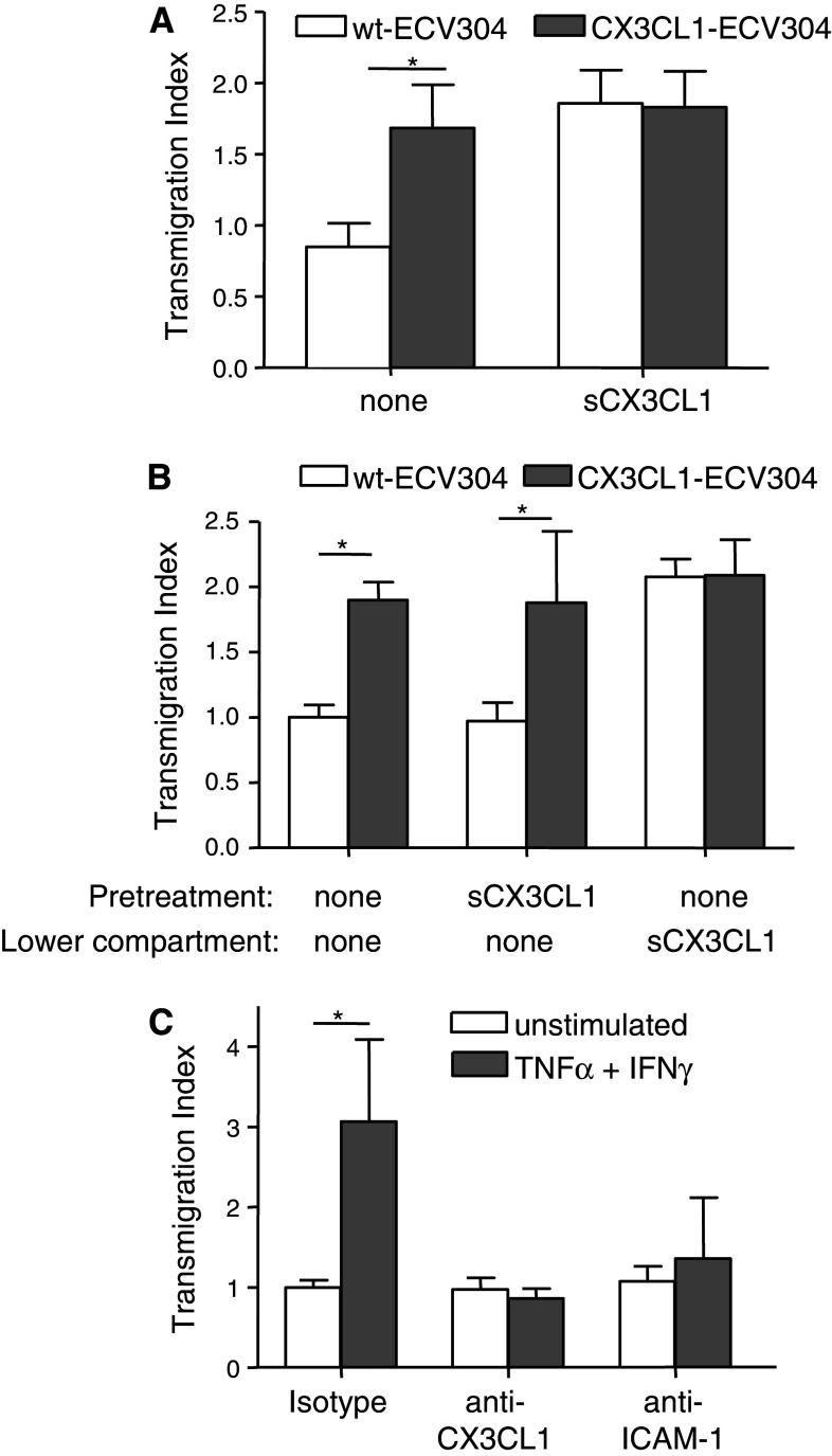
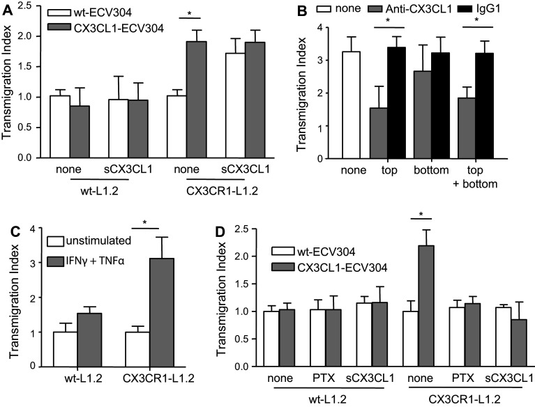
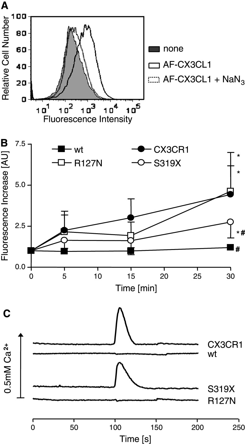
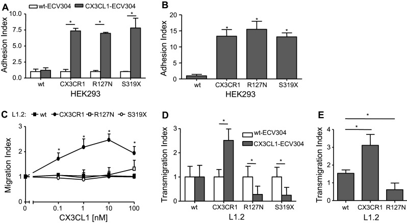
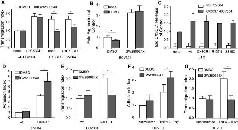
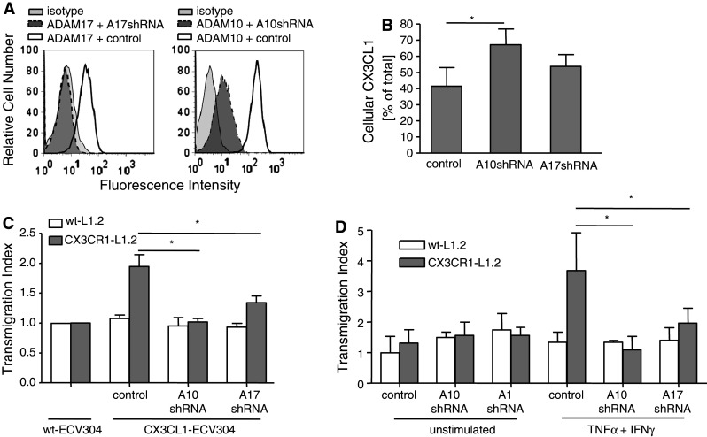
The surface-expressed transmembrane CX3C chemokine ligand 1 (CX3CL1/fractalkine) induces firm adhesion of leukocytes expressing its receptor CX3CR1. After shedding by the disintegrins and metalloproteinases (ADAM) 10 and 17, CX3CL1 also acts as soluble leukocyte chemoattractant. Here, we demonstrate that transmembrane CX3CL1 expressed on both endothelial and epithelial cells induces leukocyte transmigration. To investigate the underlying mechanism, we generated CX3CR1 variants lacking the intracellular aspartate-arginine-tyrosine (DRY) motif or the intracellular C-terminus which led to a defect in intracellular calcium response and impaired ligand uptake, respectively. While both variants effectively mediated firm cell adhesion, they failed to induce transmigration and rather mediated retention of leukocytes on the CX3CL1-expressing cell layer. Targeting of ADAM10 led to increased adhesion but reduced transmigration in response to transmembrane CX3CL1, while transmigration towards soluble CX3CL1 was not affected. Thus, transmembrane CX3CL1 mediates leukocyte transmigration via the DRY motif and C-terminus of CX3CR1 and the activity of ADAM10.


Figures

Fig. 1
Fig. 1
Transmembrane CX3CL1 promotes transmigration. a Wt- or CX3CL1-ECV304 cells were grown in transwell inserts and washed to remove free CX3CL1. As a control, recombinant soluble CX3CL1 was again added to the lower transwell compartment. Subsequently, cells were assayed for transmigration of freshly prepared PBMC that were added to the upper transwell compartment. PBMC that transmigrated into the lower compartment were counted and results were expressed as transmigration index representing the fold increase over random migration. b Wt- or CX3CL1-ECV304 cells were pretreated with recombinant soluble CX3CL1 (10 nM) that was added to the upper transwell compartment. After 1 h, cells were washed and assayed for transmigration of PBMC in the absence or presence of recombinant soluble CX3CL1 in the lower compartment. c HUVEC were left unstimulated or stimulated with IFN-γ and TNF-α (both 10 ng/ml) for 16 h and subsequently treated with neutralizing antibodies against CX3CL1 and ICAM-1 or isotype control (10 μg/ml each) for 1 h. After washing to remove free antibody, HUVEC were transferred into fresh medium and assayed for PBMC transmigration. Data are shown as mean plus SD of three independent experiments. The asterisks indicate statistically significant differences versus the controls (untransfected, unstimulated or isotype, respectively, p < 0.05)
Fig. 2
Fig. 2
CX3CR1 mediates transmigration. a Wt- and CX3CR1-L1.2 were investigated for transmigration across wt- or CX3CL1-ECV304 cells cultured in transwell inserts. b Neutralizing monoclonal antibody to CX3CL1 or isotype control (10 μg/ml each) was added to the lower or upper compartment of transwells containing wt- or CX3CL1-ECV304 cells. After 15 min, the antibody was removed and the cells were investigated for transmigration of CX3CR1-L1.2 cells. c HUVEC were stimulated with IFN-γ and TNF-α (both 10 ng/ml) for 16 h or left untreated. Subsequently, wt- and CX3CR1-L1.2 cells were assayed for transmigration across the HUVEC layer. d Wt- and CX3CR1-L1.2 cells were pretreated for 30 min with soluble CX3CL1 (10 nM) or 2 h with PTX (100 ng/ml) and subsequently assayed for transmigration across wt- or CX3CL1-ECV304 cells. Data are shown as mean plus SD of three independent experiments. The asterisks indicate statistically significant differences versus the controls (untransfected, unstimulated or isotype, respectively, p < 0.05)
Fig. 3
Fig. 3
Design and expression of CX3CR1 mutants. a CX3CR1 carries a number of conserved sequences including a DRY motif in the second intracellular loop, a NPX2-3Y motif in the seventh transmembrane domain and several serine residues at the C-terminus. To disrupt these motifs, CX3CR1 was mutated at the indicated sites. R127 in the second intracellular loop was mutated to N (R127N), N289 and Y293 in the seventh transmembrane domain were changed into A (N289A and Y293A, respectively), and the intracellular C-terminus was truncated before S319 (S319X). b Expression of the receptor variants was controlled by flow cytometry using PE-conjugated rat anti-CX3CR1. Ligand binding was analyzed by flow cytometry using a recombinant CX3CL1-Fc construct as a ligand. Receptor expression and ligand binding were measured as the mean fluorescence intensity increase compared to control. After logarithmic transformation, the data were summarized as means plus SD from five independent experiments. Expression in mock cells was used as a covariate. The asterisks indicate statistically significant differences (p < 0.05) in receptor expression and ligand binding between CX3CR1 and its variants. c CX3CR1 and its R127N and S319X variants were then stably expressed in HEK293 cells and selected for comparable surface expression by flow cytometry using PE-conjugated rat anti-CX3CR1. Cells were constantly controlled for stable receptor expression. Data are shown as a representative histogram
Fig. 4
Fig. 4
Ligand uptake and calcium signaling via CX3CR1 mutants. a HEK293 cells expressing the different receptor variants were incubated with AlexaFluor647-labeled CX3CL1 (AF-CX3CL1) at 37°C in the absence or presence of NaN3 (0.2%). Subsequently, cells were analyzed for binding and uptake of fluorescent CX3CL1 by flow cytometry. A representative histogram of the fluorescence signal for CX3CR1-HEK293 cells is shown. b HEK293 cells expressing the different receptor variants were incubated with AF-CX3CL1 at 37°C for the time periods indicated. The fluorescence intensity for the different HEK293 mutants was expressed in relation to that of the control receiving no AF-CX3CL1 and is presented as mean plus SD from three independent experiments. The asterisks indicate statistically significant differences versus the untransfected control. The crosses indicate statistically significant differences versus CX3CR1-HEK293 cells (p < 0.05). c HEK293 cells expressing the indicated receptor variants were loaded with calcium indicator Fluo-3-AM and changes in fluorescence intensity upon treatment with soluble CX3CL1 (10 nM at 100 s) were recorded. The arrow indicates an increase of 0.5 mM Ca2+. Results shown are representative for three independent experiments
Fig. 5
Fig. 5
Role of the DRY motif and the C-terminus for CX3CR1-mediated adhesion and transmigration. The ability of the receptor variants to mediate adhesion to CX3CL1 was tested under static (a) and flow (b) conditions. a For static adhesion assays, calcein-labeled HEK293 cells expressing the indicated receptor variants were seeded onto wt- or CX3CL1-ECV304 cells. After washing, the fluorescence signal of the adhering cells was measured. The adhesion index was determined by calculating the ratio of the fluorescence signal from adherent HEK293 cells bound to CX3CL1-ECV304 and from that bound to wt-ECV304 cells. b For flow adhesion experiments, labeled HEK293 cells expressing the indicated receptor variants were perfused over a layer of cytokine-stimulated HUVEC for 1 min. c L1.2 cells expressing the indicated receptor variants were tested for their chemotactic response towards increasing concentrations of soluble CX3CL1 (0.1–100 nM) in a modified Boyden chamber assay. d L1.2 cells expressing the indicated receptor variants were studied for transmigration through CX3CL1-ECV304 cells. e HUVEC were stimulated with IFN-γ and TNF-α (both 10 ng/ml) for 16 h and subsequently probed for transmigration of L1.2 cells in response to CX3CR1 or its R127N variant. Three independent experiments were performed and data are shown as mean plus SD. The asterisks indicate statistically significant differences versus the untransfected control (p < 0.05)
Fig. 6
Fig. 6
Effect of the metalloproteinase inhibitor GW280264X on CX3CL1-mediated transmigration. a Wt- or CX3CL1-ECV304 cells were grown on transwell inserts and pretreated with GW280264X (10 μM) or DMSO control for 1 h. After removal of the inhibitor, the lower compartments of the transwell systems received soluble CX3CL1 (10 nM) or were left without stimulus. Subsequently, cells were assayed for transmigration of wt- and CX3CR1-L1.2 cells. b CX3CL1-ECV304 cells were pretreated with GW280264X (10 μM) or DMSO for 1 h, washed and co-incubated with freshly prepared PBMC for 3 h. Cells were harvested and analyzed for CX3CL1 surface expression by flow cytometry. Surface expression was expressed in relation to that of CX3CL1-ECV304 cells receiving no inhibitor and no PBMC. c Wt- or CX3CL1-ECV304 cells were incubated for 3 h in the presence or absence of L1.2 cells expressing the indicated CX3CR1 variants and subsequently conditioned media were analyzed for the presence of shed soluble CX3CL1 by ELISA. Results are expressed in relation to the control receiving no L1.2 cells. d Wt- or CX3CL1-ECV304 cells were pretreated with DMSO control or GW280264X (10 μM) for 1 h, washed and subsequently analyzed for adhesion of PBMC under flow. e Wt- or CX3CL1-ECV304 cells were grown on transwell inserts, pretreated with DMSO control or GW280264X (10 μM) for 1 h, and subsequently probed for transmigration of PBMC. f HUVEC were stimulated with IFN-γ and TNF-α (both 10 ng/ml) or left unstimulated for 16 h. Subsequently, cells were pretreated with DMSO control or GW280264X (10 μM) for 1 h and then analyzed for flow-resistant adhesion of PBMC. g Unstimulated and IFN-γ/TNF-α-stimulated HUVEC were pretreated with DMSO control or GW280264X and subsequently probed for transmigration of PBMC. Data are shown as mean plus SD obtained from three independent experiments. Statistically significant differences between inhibitor-treated and DMSO-treated cells are indicated by asterisks (p < 0.05)
Fig. 7
Fig. 7
Role of ADAM10 and ADAM17 for CX3CL1-induced transmigration. Wt- and CX3CL1-ECV304 cells were stably transduced with lentivirus carrying shRNA for knockdown of ADAM10 and ADAM17 (A10shRNA or A17shRNA) or scrambled shRNA control, respectively. a Efficient and sustained downregulation of ADAM10 and ADAM17 surface expression was controlled by flow cytometry and is shown as representative histograms. b Stably transduced CX3CL1-ECV304 cells were incubated for 2 h, and subsequently released CX3CL1 in the supernatants and cell-associated CX3CL1 in the lysates were quantified by ELISA. The amount of residual cell-expressed CX3CL1 was calculated in relation to the total content of CX3CL1 in both the lysates and supernatants. c Stably transduced CX3CL1-ECV304 cells were investigated for transmigration by wt- and CX3CR1-L1.2 cells. d HUVEC were transduced with A10shRNA, A17shRNA or scramble shRNA control. Cells were stimulated with IFN-γ and TNF-α (both 10 ng/ml) or left unstimulated for 16 h and subsequently investigated for transmigration by wt- and CX3CR1-L1.2 cells. Data are shown as mean plus SD obtained from three independent experiments. Statistically significant differences versus the controls receiving scrambled shRNA are indicated by asterisks (p < 0.05)

References

    1. Springer TA. Traffic signals for lymphocyte recirculation and leukocyte emigration: the multistep paradigm. Cell. 1994;76:301–314. doi: 10.1016/0092-8674(94)90337-9. - DOI - PubMed
    1. Combadiere C, Potteaux S, Gao JL, Esposito B, Casanova S, Lee EJ, Debre P, Tedgui A, Murphy PM, Mallat Z. Decreased atherosclerotic lesion formation in CX3CR1/apolipoprotein E double knockout mice. Circulation. 2003;107:1009–1016. doi: 10.1161/01.CIR.0000057548.68243.42. - DOI - PubMed
    1. Lesnik P, Haskell CA, Charo IF. Decreased atherosclerosis in CX3CR1−/− mice reveals a role for fractalkine in atherogenesis. J Clin Invest. 2003;111:333–340. - PMC - PubMed
    1. Teupser D, Pavlides S, Tan M, Gutierrez-Ramos JC, Kolbeck R, Breslow JL. Major reduction of atherosclerosis in fractalkine (CX3CL1)-deficient mice is at the brachiocephalic artery, not the aortic root. Proc Natl Acad Sci USA. 2004;101:17795–17800. doi: 10.1073/pnas.0408096101. - DOI - PMC - PubMed
    1. McDermott DH, Fong AM, Yang Q, Sechler JM, Cupples LA, Merrell MN, Wilson PW, D’Agostino RB, O’Donnell CJ, Patel DD, Murphy PM. Chemokine receptor mutant CX3CR1–M280 has impaired adhesive function and correlates with protection from cardiovascular disease in humans. J Clin Invest. 2003;111:1241–1250. - PMC - PubMed

Publication types

MeSH terms