Review
. 2010 Aug;3(4):374-83.
doi: 10.1007/s12265-010-9174-x. Epub 2010 Mar 16.

Mitochondrial pruning by Nix and BNip3: an essential function for cardiac-expressed death factors

Affiliations
Review

Mitochondrial pruning by Nix and BNip3: an essential function for cardiac-expressed death factors

Gerald W Dorn 2nd. J Cardiovasc Transl Res. 2010 Aug.

Abstract

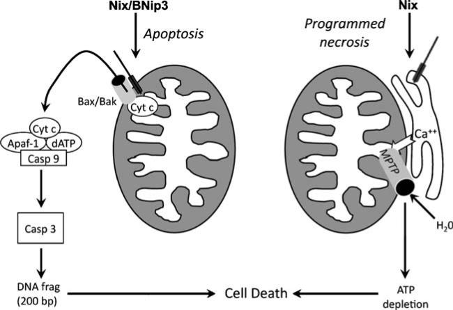
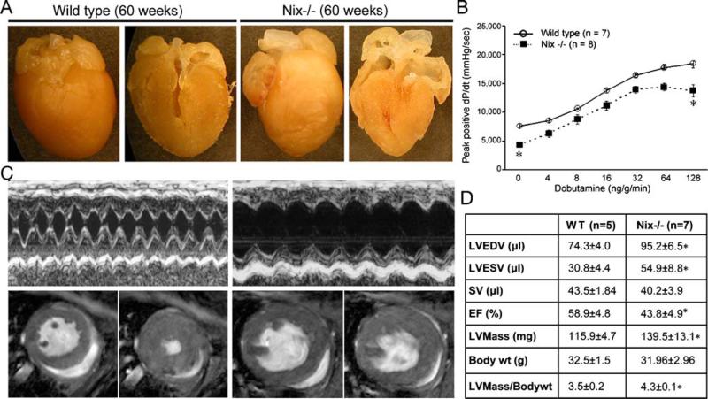
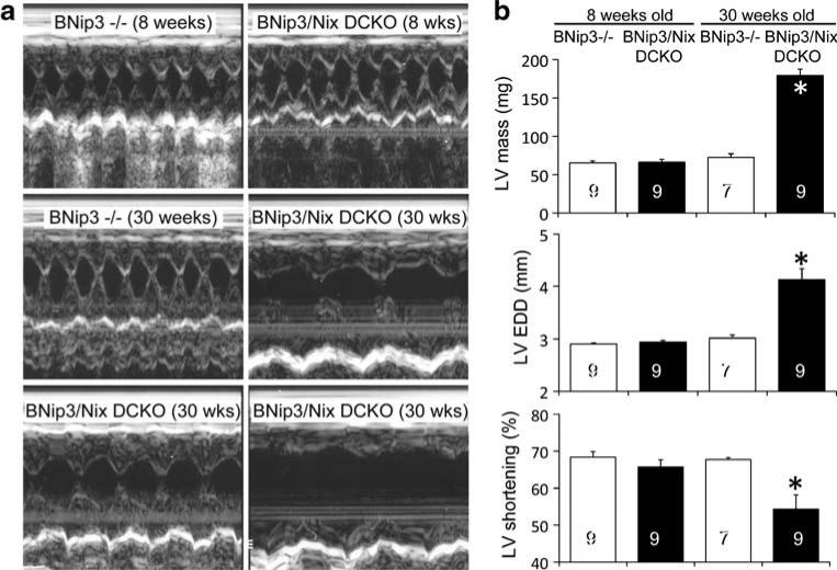
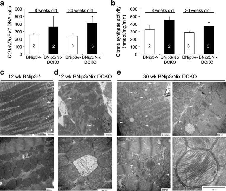
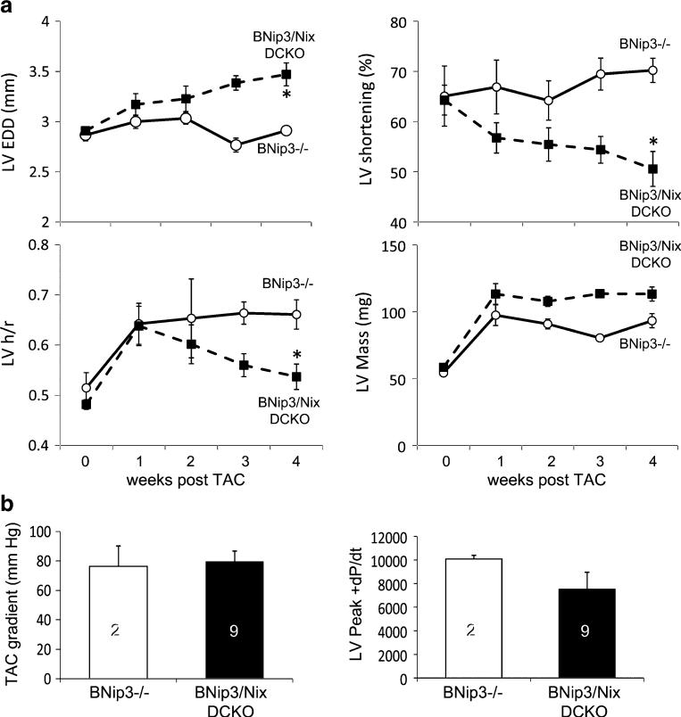
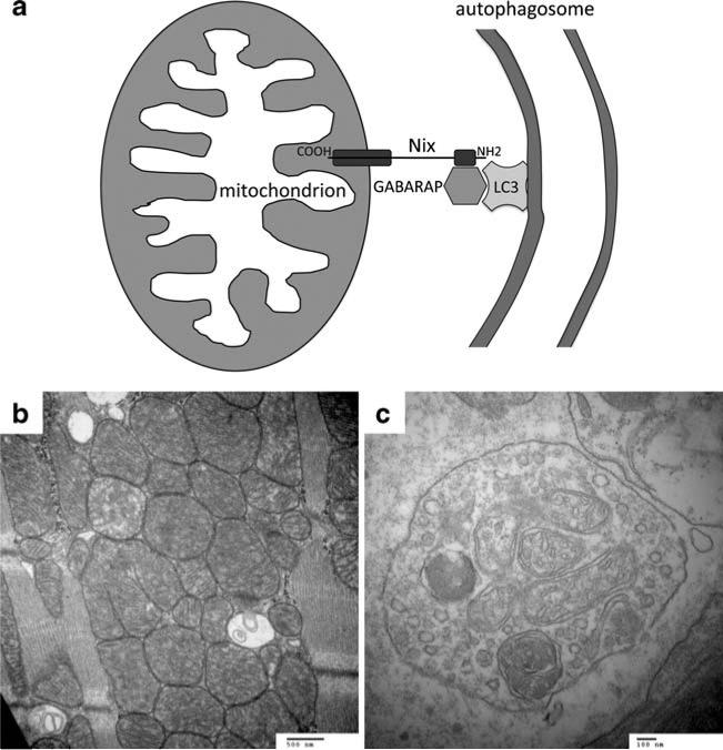
Programmed cardiac myocyte death via the intrinsic, or mitochondrial, pathway is a mechanism of pathological ventricular remodeling after myocardial infarction and during chronic pressure overload hypertrophy. Transcriptional upregulation of the closely related proapoptotic Bcl2 family members BNip3 in ischemic myocardium and Nix in hypertrophied myocardium suggested a molecular mechanism by which programmed cell death can be initiated by cardiac stress and lead to dilated cardiomyopathy. Studies using transgenic and gene knockout mice subsequently demonstrated that expression of BNip3 and Nix is both sufficient for cardiomyopathy development and necessary for cardiac remodeling after reversible coronary occlusion and transverse aortic banding, respectively. Here, these data are reviewed in the context of recent findings showing that Nix not only stimulates cardiomyocyte apoptosis but also induces mitochondrial autophagy (mitophagy) and indirectly activates the mitochondrial permeability transition pore, causing cell necrosis. New findings are presented suggesting that Nix and BNip3 have an essential function, "mitochondrial pruning," that restrains mitochondrial proliferation in cardiomyocytes and without which an age-dependent mitochondrial cardiomyopathy develops.


Figures

Fig. 1
Fig. 1
Schematic diagram depicting canonical apoptotic function of Nix and BNip3 and atypical MTPT opening by Nix. Left, Nix and BNip3 localize to mitochondrial outer membranes via carboxyl-terminal hydrophobic domains and stimulate membrane permeabilization by Bax and/or Bak. Cytochrome c released into the cytoplasm binds in a functional “apoptosome” with other factors to activate initiator caspase 9, resulting in cascade activation of effector caspase 3, DNA fragmentation, and apoptosis. Right, Nix localizes to endoplasmic and sarcoplasmic reticulum where it increases reticular calcium via a poorly defined mechanism. Reticular calcium transferred to mitochondria through calcium “hot spots” is takenupbythe mitochondrial calcium uniporter and opens mitochondrial permeability transition pores, resulting in osmotic swelling and mitochondrial rupture. Although cytochrome c is released by this mechanism, cell death occurs because of metabolic shutdown, not apoptosis
Fig. 2
Fig. 2
Cardiomegaly and contractile depression in 60-week-old Nix knockout mice. a Images of intact (left panels) and halved (right panels) hearts from 60-week-old wild-type and germ-line Nix knockout (Nix−/−). b Invasive hemodynamic assessment of isovolumic contractility as a function of dobutamine dose in 60-week-old mice. c Representative M-mode echocardiograms (top) and MRI (bottom) demonstrating left ventricular enlargement and diminished ejection performance but absence of wall thinning. For MRI, left panels are diastole, right panels are systole. d Quantitative morpho-metric and functional data from MRI studies. LVEDV left ventricular end diastolic volume, LVESD left ventricular end systolic volume, SV stroke volume, EF ejection fraction
Fig. 3
Fig. 3
Age-dependent cardiomyopathy in 30-week-old Nix/BNip3 double cardiac knockout (DCKO) mice. a Representative M-mode echocardiograms from 8-week BNip3−/− and DCKO mice (top), from a 30-week BNip3−/− mouse (middle, left), and from three 30-week DCKO mice (middle, right, and bottom). b Quantitative group echocardiographic data. LVEDD left ventricular end diastolic dimension
Fig. 4
Fig. 4
Mitochondrial abnormalities in Nix/BNip3 double cardiac knockout mice. a Quantitative PCR of cardiac mitochondrial CO1 and nuclear NDUFV1 gene content. b Myocardial citrate synthase activity. c–e Transmission EM of myocardial samples from different genotypes and ages: c Twelve-week-old BNip3−/−. Top, 6,000×; bottom, 30,000×. d Twelve-week-old DCKO. Top, 6,000×; bottom, 70,000×. e Thirty-week-old DCKO. Top left, 6,000×; top right, 25,000×; bottom left, 30,000×; bottom right, 50,000×
Fig. 5
Fig. 5
Rapid decompensation of Nix/BNip3 double cardiac knockout (DCKO) mice after acute pressure overloading. a Time-dependent changes in echocardiographically determined left ventricular end diastolic dimension (LVEDD, upper left), fractional shortening (upper right), ratio of wall thickness to radius (h/r, bottom left), and mass (lower right) after transverse aortic constriction (TAC). b Transverse aortic gradient (left) and peak positive dP/dt (right) at terminal invasive hemodynamic studies 4 weeks after TAC
Fig. 6
Fig. 6
Schematic diagram depicting proposed molecular mechanism for mitochondrial pruning by Nix and BNip3. a Nix localizes to mitochondrial outer membranes via hydrophobic C-terminal domain (COOH) and to GABARAP via amino terminal domain (NH2). GABARAP acts as a docking protein with autophagic membrane-associated LC3 to target Nix-labeled mitochondrion for autophagic clearance. b Mitochondrial replaced by double membrane delimited autophagic vesicles in a senescent BNip3 knockout mouse heart (30,000×). c High magnification (80,000×) of autophagic vesicle containing mitochondrial remnants

Similar articles

Cited by

References

    1. Diwan A, Dorn GW., II Decompensation of cardiac hypertrophy: Cellular mechanisms and novel therapeutic targets. Physiology (Bethesda) 2007;22:56–64. - PubMed
    1. Berry JJ, Hoffman JM, Steenbergen C, Baker JA, Floyd C, Van Trigt P, et al. Human pathologic correlation with PET in ischemic and nonischemic cardiomyopathy. Journal of Nuclear Medicine. 1993;34:39–47. - PubMed
    1. Olivetti G, Abbi R, Quaini F, Kajstura J, Cheng W, Nitahara JA, et al. Apoptosis in the failing human heart. Journal of Nuclear Medicine. 1997;336:1131–1141. - PubMed
    1. Francis GS. Changing the remodeling process in heart failure: Basic mechanisms and laboratory results. Current Opinion in Cardiology. 1998;13:156–161. - PubMed
    1. Olivetti G, Capasso JM, Sonnenblick EH, Anversa P. Side-to-side slippage of myocytes participates in ventricular wall remodeling acutely after myocardial infarction in rats. Circulation Research. 1990;67:23–34. - PubMed

Publication types