Review
. 2010 Nov;20(11):2707-14.
doi: 10.1007/s00330-010-1845-0. Epub 2010 Jun 18.

Radiation exposure in X-ray-based imaging techniques used in osteoporosis

Affiliations
Review

Radiation exposure in X-ray-based imaging techniques used in osteoporosis

John Damilakis et al. Eur Radiol. 2010 Nov.

Abstract

Recent advances in medical X-ray imaging have enabled the development of new techniques capable of assessing not only bone quantity but also structure. This article provides (a) a brief review of the current X-ray methods used for quantitative assessment of the skeleton, (b) data on the levels of radiation exposure associated with these methods and (c) information about radiation safety issues. Radiation doses associated with dual-energy X-ray absorptiometry are very low. However, as with any X-ray imaging technique, each particular examination must always be clinically justified. When an examination is justified, the emphasis must be on dose optimisation of imaging protocols. Dose optimisation is more important for paediatric examinations because children are more vulnerable to radiation than adults. Methods based on multi-detector CT (MDCT) are associated with higher radiation doses. New 3D volumetric hip and spine quantitative computed tomography (QCT) techniques and high-resolution MDCT for evaluation of bone structure deliver doses to patients from 1 to 3 mSv. Low-dose protocols are needed to reduce radiation exposure from these methods and minimise associated health risks.


Figures
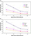
Fig. 1
Fig. 1
Effective doses from a single DXA of the spine (a) or hip (b) as a function of patient age (adapted from Blake et al. [33]). Patient dose was estimated for Hologic DXA using three imaging modes: Array mode (60-s data acquisition time); Fast mode (30-s data acquisition time); and Express mode (10-s data acquisition time)

Similar articles

Cited by

References

    1. Compston JE, Papapoulos SE, Blanchard F. Report on osteoporosis in the European Community: current status and recommendations for the future. Osteoporos Int. 1998;8:531–534. doi: 10.1007/s001980050094. - DOI - PubMed
    1. Gehlbach SH, Bigelow C, Heimisdottir M, et al. Recognition of vertebral fracture in a clinical setting. Osteoporos Int. 2000;11:577–582. doi: 10.1007/s001980070078. - DOI - PubMed
    1. Genant HK, Wu CY, van Kuijk C, Nevitt MC. Vertebral fracture assessment using a semiquantitative technique. J Bone Miner Res. 1993;8:1137–1148. doi: 10.1002/jbmr.5650080915. - DOI - PubMed
    1. Jiang G, Eastell R, Barrington NA, Ferrar L. Comparison of methods for the visual identification of prevalent vertebral fracture in osteoporosis. Osteoporos Int. 2004;15:887–896. doi: 10.1007/s00198-004-1626-1. - DOI - PubMed
    1. Guglielmi G, Diacinti D, van Kuijk C, et al. Vertebral morphometry: current methods and recent advances. Eur Radiol. 2008;18:1484–1496. doi: 10.1007/s00330-008-0899-8. - DOI - PubMed