. 2010 Jun;26(6):717-23.
doi: 10.1089/aid.2009.0254.

Structural conservation predominates over sequence variability in the crown of HIV type 1's V3 loop

Affiliations

Structural conservation predominates over sequence variability in the crown of HIV type 1's V3 loop

David Almond et al. AIDS Res Hum Retroviruses. 2010 Jun.

Abstract

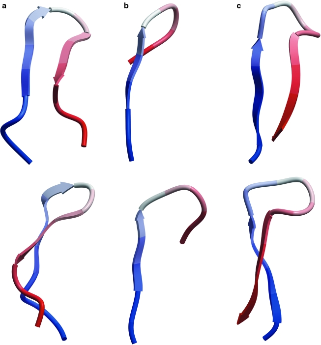
The diversity of HIV-1 is a confounding problem for vaccine design, as the human immune response appears to favor poor or strain-specific responses to any given HIV-1 virus strain. A significant portion of this diversity is manifested as sequence variability in the loops of HIV-1's surface envelope glycoprotein. Here we show that the most variable sequence positions in the third variable (V3) loop crown cluster to a small zone on the surface of one face of the V3 loop ss-hairpin conformation. These results provide a novel visualization of the gp120 V3 loop, specifically demonstrating a surprising preponderance of conserved three-dimensional structure in a highly sequence-variable region. From a structural point of view, there appears to be less diversity in this region of the HIV-1 "principle neutralizing domain" than previously appreciated.


Figures

FIG. 1.
FIG. 1.
Ribbon diagrams of three HIV gp120 V3 loops complexed with different anti-V3 mAbs, as viewed from the front (top) and side (bottom). Thus, V3 loops are shown in the context of (a) mAb 2219, (b) mAb 447-52D, and (c) mAb F425-B4e8. Loops a and b are from HIV-1 strain MN, whereas c is from strain RP142. Each 3D structure is colored from N-terminal to C-terminal in a blue to red gradient.
FIG. 2.
FIG. 2.
(a) Plot of the occurrence of the tetrameric motif in the V3 loop crown sequence (at the ß-turn location) in a representative sample of all known V3 loops. Of the recorded motifs occurring in the turn sequence 94% have already been identified in the literature as sequences that strongly predict a ß-turn or ß-hairpin conformation. Of the remaining 6%, 5% are sequences that moderately predict a ß-turn and none is a strongly α-helical sequence. (b) Design and testing of a V3 loop with an α-helical crown (designated S2, bottom panel). The ribbon structure shown below each sequence is the ab initio folding of the red or green colored portion of the sequence, a technique previously shown to predict the structural preferences of the V3 loop crown accurately enough to recapitulate crystallographic V3 loop conformations. The upper sequence shown in green is the wild-type Hxbc2 V3 crown sequence (HXB2WT). The lower sequence differs only in the amino acids shown in red, which are designed to fold ab initio an α-helical V3 loop sequence (HX S2). (c) Infectivity of HXB2 wild-type (HXB2 WT) and V3 loop engineered HXB2 chimeric pseudotyped (HX S2) viruses. The result shows mean luciferase activity (y-axis) resulting from productive infection of cells with virus ± SD from triplicate wells. The α-helical V3 crown construct HX S2 shows no activity and thus is not infective, whereas HXB2WT shows the full luciferase activity typical of productive infection of cells by virus.
FIG. 3.
FIG. 3.
Average sequence variability of V3 loop positions mapped onto the 3D structure of the 2219 mAb-bound conformation of the crown of the V3 loop of clade B strain RP322 consisting of residues 8–23 of the V3 loop 8KRKRIHIGPGRAFYTT. The V3 loop crown region is divided into four different zones: the left-most division (orange) contains the base of the V3 crown (8KRK and 23T). The upper middle division (red) contains the residues pointing their side chains into the hydrophilic face of the amphipathic ß-hairpin (11R, 13H, 19A and 22T). The lower middle division (blue) contains residues that point their side chains into the hydrophobic face of the amphipathic ß-hairpin (12I, 14I, 20F and 21Y). The division on the right (green) contains the conserved ß-turn (15GPGR). The average sequence variability score, calculated as noted in Materials and Methods, is noted in each region.

References

    1. Huang C. Tang M. Zhang M, et al. Structure of a V3-containing HIV-1 gp120 core. Science. 2005;310(5750):1025–1028. - PMC - PubMed
    1. Lo Conte L. Ailey B. Hubbard T. Brenner S. Murzin A. Chothia C. SCOP: A structural classification of proteins database. Nucleic Acids Res. 2000;28(1):257–259. - PMC - PubMed
    1. Trkola A. Dragic T. Arthos J, et al. CD4-dependent, antibody-sensitive interactions between HIV-1 and its co-receptor CCR-5. Nature. 1996;384(6605):184–187. - PubMed
    1. Hill C. Deng H. Unutmaz D, et al. Envelope glycoproteins from human immunodeficiency virus types 1 and 2 and simian immunodeficiency virus can use human CCR5 as a coreceptor for viral entry and make direct CD4-dependent interactions with this chemokine receptor. J Virol. 1997;71(9):6296–6304. - PMC - PubMed
    1. Leitner T, editor; Foley B, editor; Hahn B, et al., editors. HIV Sequence Compendium. Los Alamos National Laboratory, NM: Theoretical Biology and Biophysics Group; 2005. No. LA-UR 06-0680.

Publication types

Substances