

Taxol, a microtubule stabilizer, prevents ischemic ventricular arrhythmias in rats
- PMID: 20561109
- PMCID: PMC3822629
- DOI: 10.1111/j.1582-4934.2010.01106.x
Taxol, a microtubule stabilizer, prevents ischemic ventricular arrhythmias in rats
Abstract
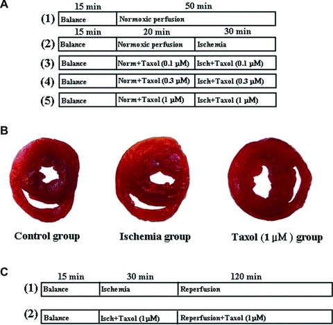
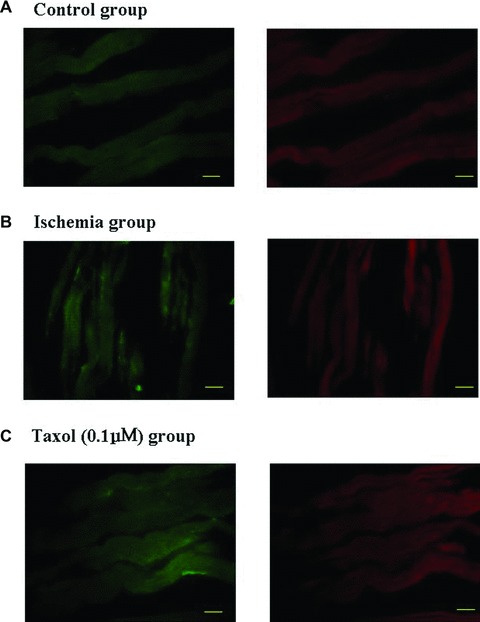
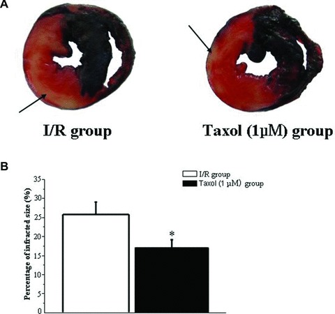
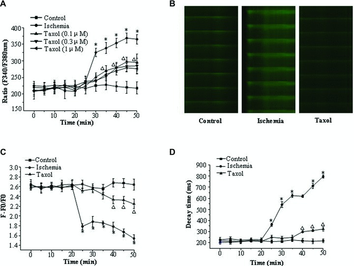
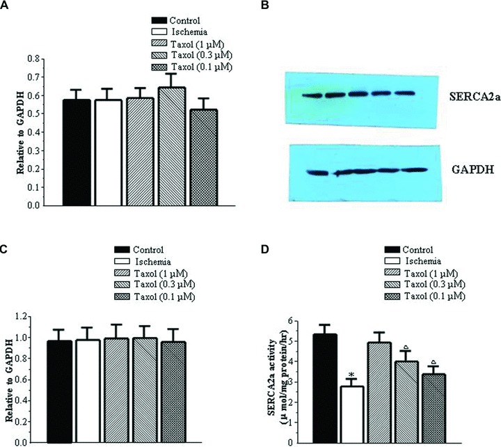
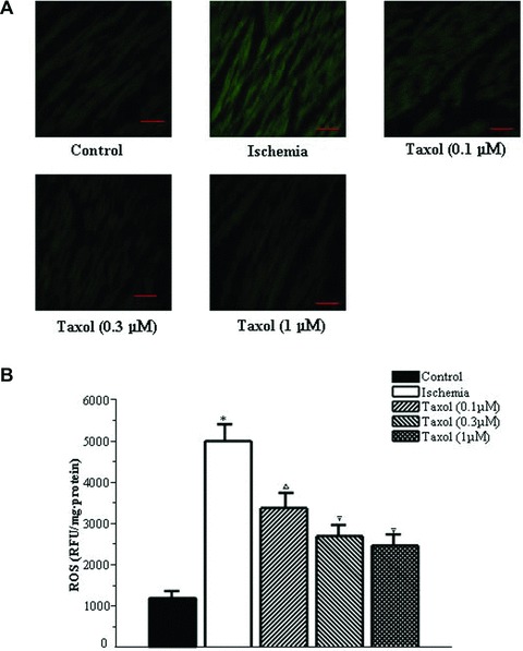
Microtubule integrity is important in cardio-protection, and microtubule disruption has been implicated in the response to ischemia in cardiac myocytes. However, the effects of Taxol, a common microtubule stabilizer, are still unknown in ischemic ventricular arrhythmias. The arrhythmia model was established in isolated rat hearts by regional ischemia, and myocardial infarction model by ischemia/reperfusion. Microtubule structure was immunohistochemically measured. The potential mechanisms were studied by measuring reactive oxygen species (ROS), activities of oxidative enzymes, intracellular calcium concentration ([Ca(2+) ](i) ) and Ca(2+) transients by using fluorometric determination, spectrophotometric assays and Fura-2-AM and Fluo-3-AM, respectively. The expression and activity of sarcoplasmic reticulum Ca(2+)-ATPase (SERCA2a) was also examined using real-time polymerase chain reaction, Western blot and pyruvate/Nicotinamide adenine dinucleotide-coupled reaction. Our data showed that Taxol (0.1, 0.3 and 1 μM) effectively reduced the number of ventricular premature beats and the incidence and duration of ventricular tachycardia. The infarct size was also significantly reduced by Taxol (1 μM). At the same time, Taxol preserved the microtubule structure, increased the activity of mitochondrial electron transport chain complexes I and III, reduced ROS levels, decreased the rise in [Ca(2+)](i) and preserved the amplitude and decay times of Ca(2+) transients during ischemia. In addition, SERCA2a activity was preserved by Taxol during ischemia. In summary, Taxol prevents ischemic ventricular arrhythmias likely through ameliorating abnormal calcium homeostasis and decreasing the level of ROS. This study presents evidence that Taxol may be a potential novel therapy for ischemic ventricular arrhythmias.
© 2011 The Authors Journal of Cellular and Molecular Medicine © 2011 Foundation for Cellular and Molecular Medicine/Blackwell Publishing Ltd.
Figures
Similar articles
-
Taxol, a microtubule stabilizer, improves cardiac functional recovery during postischemic reperfusion in rat in vitro.Cardiovasc Ther. 2012 Feb;30(1):12-30. doi: 10.1111/j.1755-5922.2010.00163.x. Epub 2010 Jun 14. Cardiovasc Ther. 2012. PMID: 20553295
-
Taxol, a microtubule stabilizer, improves cardiac contractile function during ischemia in vitro.Pharmacology. 2010;85(5):301-10. doi: 10.1159/000292948. Epub 2010 May 7. Pharmacology. 2010. PMID: 20453554
-
Taxol prevents myocardial ischemia-reperfusion injury by inducing JNK-mediated HO-1 expression.Pharm Biol. 2016;54(3):555-60. doi: 10.3109/13880209.2015.1060507. Epub 2015 Aug 13. Pharm Biol. 2016. PMID: 26270131
-
Microtubule stabilization with paclitaxel does not protect against infarction in isolated rat hearts.Exp Physiol. 2015 Jan;100(1):23-34. doi: 10.1113/expphysiol.2014.082925. Epub 2014 Dec 9. Exp Physiol. 2015. PMID: 25557728
-
Expression of SERCA isoform with faster Ca2+ transport properties improves postischemic cardiac function and Ca2+ handling and decreases myocardial infarction.Am J Physiol Heart Circ Physiol. 2007 Oct;293(4):H2418-28. doi: 10.1152/ajpheart.00663.2007. Epub 2007 Jul 13. Am J Physiol Heart Circ Physiol. 2007. PMID: 17630344
Cited by
-
Single-nucleotide variations in cardiac arrhythmias: prospects for genomics and proteomics based biomarker discovery and diagnostics.Genes (Basel). 2014 Mar 27;5(2):254-69. doi: 10.3390/genes5020254. Genes (Basel). 2014. PMID: 24705329 Free PMC article.
-
Farnesyl pyrophosphate synthase inhibitor, ibandronate, improves endothelial function in spontaneously hypertensive rats.Mol Med Rep. 2016 May;13(5):3787-96. doi: 10.3892/mmr.2016.5025. Epub 2016 Mar 21. Mol Med Rep. 2016. PMID: 27035426 Free PMC article.
-
Role of Ran-regulated nuclear-cytoplasmic trafficking of pVHL in the regulation of microtubular stability-mediated HIF-1α in hypoxic cardiomyocytes.Sci Rep. 2015 Mar 17;5:9193. doi: 10.1038/srep09193. Sci Rep. 2015. PMID: 25779090 Free PMC article.
-
Microtubular stability affects pVHL-mediated regulation of HIF-1alpha via the p38/MAPK pathway in hypoxic cardiomyocytes.PLoS One. 2012;7(4):e35017. doi: 10.1371/journal.pone.0035017. Epub 2012 Apr 10. PLoS One. 2012. PMID: 22506063 Free PMC article.
-
Identification of cardioprotective drugs by medium-scale in vivo pharmacological screening on a Drosophila cardiac model of Friedreich's ataxia.Dis Model Mech. 2018 Jul 20;11(7):dmm033811. doi: 10.1242/dmm.033811. Dis Model Mech. 2018. PMID: 29898895 Free PMC article.
References
-
- Prunier F, Kawase Y, Gianni D, et al. Prevention of ventricular arrhythmias with sarcoplasmic reticulum Ca2+ ATPase pump overexpression in a porcine model of ischemia reperfusion. Circulation. 2008;118:614–24. - PubMed
-
- Members of the Sicilian G. New approaches to antiarrhythmic therapy: emerging therapeutic applications of the cell biology of cardiac arrhythmias. Circulation. 2001;104:345–60. - PubMed
-
- Gomez AM, Kerfant BG, Vassort G. Microtubule disruption modulates Ca2+ signaling in rat cardiac myocytes. Circ Res. 2000;86:30–6. - PubMed
Publication types
MeSH terms
Substances
LinkOut - more resources
Full Text Sources
Medical
Miscellaneous
