Review
. 2010 Jul;21(3):511-27.
doi: 10.1016/j.nec.2010.03.006.

Cerebral venous sinus (sinovenous) thrombosis in children

Affiliations
Review

Cerebral venous sinus (sinovenous) thrombosis in children

Nomazulu Dlamini et al. Neurosurg Clin N Am. 2010 Jul.

Abstract

Cerebral venous sinus (sinovenous) thrombosis (CSVT) in childhood is a rare, but underrecognized, disorder, typically of multifactorial etiology, with neurologic sequelae apparent in up to 40% of survivors and mortality approaching 10%. There is an expanding spectrum of perinatal brain injury associated with neonatal CSVT. Although there is considerable overlap in risk factors for CSVT in neonates and older infants and children, specific differences exist between the groups. Clinical symptoms are frequently nonspecific, which may obscure the diagnosis and delay treatment. While morbidity and mortality are significant, CSVT recurs less commonly than arterial ischemic stroke in children. Appropriate management may reduce the risk of recurrence and improve outcome, however there are no randomized controlled trials to support the use of anticoagulation in children. Although commonly employed in many centers, this practice remains controversial, highlighting the continued need for high-quality studies. This article reviews the literature pertaining to pediatric venous sinus thrombosis.


Figures

Fig. 1
Fig. 1
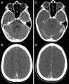
Case synopsis. A previously healthy 8-year-old girl was admitted with a 3-week history of, intermittent emesis and a 4-day history of occipital headache, and photophobia. Examination revealed severe dehydration, mild hypertension, and tachycardia. Extensive thrombosis of both deep and superficial cerebral sinovenous systems was diagnosed on head CT and anticoagulation therapy was initiated. Progressive encephalopathy developed on hospital day 5, necessitating admission to the intensive care unit. Unexplained tachycardia (heart rate >200) developed on hospital day 15 and Graves disease was ultimately diagnosed (thyrotropin <0.01 mIU/L and free T4 >77.2 pmol/L.) The patient was then started on methimazole. Comprehensive prothrombotic testing uncovered a heterozygous mutation in the Factor V Leiden gene. She completed 6 months of anticoagulation with subcutaneous low molecular weight heparin. Follow-up neurologic examination revealed mild left incoordination and bilateral kinetic tremor (left > right), perhaps secondary to hemorrhagic venous infarction of the right thalamus. (A, B) Non-contrast axial head CT done at admission revealed heterogeneous attenuation within the right transverse and sigmoid sinuses (A) and posterior aspect of the superior sagittal sinus (B), suggesting acute and subacute components of the thrombus. (C, D) Contrast CT reveals filling defects within these same sinuses. (E, F) Initial axial fluid-attenuated inversion recovery (FLAIR) (E), T1 and T2 (not shown) MRI sequences as well as diffusion-weighted imaging (DWI) (F) showed normal brain parenchyma. (H, I) A repeat MRI done in the subacute period after the patient's clinical deterioration showed increased signal within the thalami bilaterally on FLAIR (H) and T2 (not shown). Corresponding areas of diffusion restriction on DWI (I) suggested venous congestion and infarction secondary to thalamostriate venous occlusion. Peripheral blooming was seen in the right thalamus on gradient echo sequences (not shown), evidence of petechial hemorrhage. (K, L) Follow-up MRI done 6 months after diagnosis showed low FLAIR (K) and T2 signal (not shown) in the right thalamus, corresponding to hemosiderin deposits from hemorrhagic infarction. DWI (L) similarly showed low signal. (G, J, M) Three-dimensional phase contrast MR venograms performed acutely (G) and subacutely (J) showed extensive sinovenous thrombosis, involving the right transverse and sigmoid sinuses (black arrow), right internal jugular vein, posterior superior and inferior sagittal sinuses, torcula, vein of Galen, basal vein of Rosenthal, and internal cerebral and thalamostriate veins. Left parietal cortical veins were also thrombosed (white arrowheads). The left transverse and sigmoid sinuses were spared (white arrow). Interval recanalization of the left internal cerebral vein and basal vein of Rosenthal was seen subacutely (J). A 2-dimensional time-of-flight MR venogram done 6 months post diagnosis (M) showed persistently absent flow within the right transverse sinus, but partially visualized flow within the right sigmoid sinus and jugular bulb (black arrow), evidence of either partial recanalization or slow flow within these sinuses. There was complete recanalization of the superior sagittal sinus, deep venous system, and left parietal cortical veins.
Fig. 1
Fig. 1
Case synopsis. A previously healthy 8-year-old girl was admitted with a 3-week history of, intermittent emesis and a 4-day history of occipital headache, and photophobia. Examination revealed severe dehydration, mild hypertension, and tachycardia. Extensive thrombosis of both deep and superficial cerebral sinovenous systems was diagnosed on head CT and anticoagulation therapy was initiated. Progressive encephalopathy developed on hospital day 5, necessitating admission to the intensive care unit. Unexplained tachycardia (heart rate >200) developed on hospital day 15 and Graves disease was ultimately diagnosed (thyrotropin <0.01 mIU/L and free T4 >77.2 pmol/L.) The patient was then started on methimazole. Comprehensive prothrombotic testing uncovered a heterozygous mutation in the Factor V Leiden gene. She completed 6 months of anticoagulation with subcutaneous low molecular weight heparin. Follow-up neurologic examination revealed mild left incoordination and bilateral kinetic tremor (left > right), perhaps secondary to hemorrhagic venous infarction of the right thalamus. (A, B) Non-contrast axial head CT done at admission revealed heterogeneous attenuation within the right transverse and sigmoid sinuses (A) and posterior aspect of the superior sagittal sinus (B), suggesting acute and subacute components of the thrombus. (C, D) Contrast CT reveals filling defects within these same sinuses. (E, F) Initial axial fluid-attenuated inversion recovery (FLAIR) (E), T1 and T2 (not shown) MRI sequences as well as diffusion-weighted imaging (DWI) (F) showed normal brain parenchyma. (H, I) A repeat MRI done in the subacute period after the patient's clinical deterioration showed increased signal within the thalami bilaterally on FLAIR (H) and T2 (not shown). Corresponding areas of diffusion restriction on DWI (I) suggested venous congestion and infarction secondary to thalamostriate venous occlusion. Peripheral blooming was seen in the right thalamus on gradient echo sequences (not shown), evidence of petechial hemorrhage. (K, L) Follow-up MRI done 6 months after diagnosis showed low FLAIR (K) and T2 signal (not shown) in the right thalamus, corresponding to hemosiderin deposits from hemorrhagic infarction. DWI (L) similarly showed low signal. (G, J, M) Three-dimensional phase contrast MR venograms performed acutely (G) and subacutely (J) showed extensive sinovenous thrombosis, involving the right transverse and sigmoid sinuses (black arrow), right internal jugular vein, posterior superior and inferior sagittal sinuses, torcula, vein of Galen, basal vein of Rosenthal, and internal cerebral and thalamostriate veins. Left parietal cortical veins were also thrombosed (white arrowheads). The left transverse and sigmoid sinuses were spared (white arrow). Interval recanalization of the left internal cerebral vein and basal vein of Rosenthal was seen subacutely (J). A 2-dimensional time-of-flight MR venogram done 6 months post diagnosis (M) showed persistently absent flow within the right transverse sinus, but partially visualized flow within the right sigmoid sinus and jugular bulb (black arrow), evidence of either partial recanalization or slow flow within these sinuses. There was complete recanalization of the superior sagittal sinus, deep venous system, and left parietal cortical veins.
Fig. 2
Fig. 2
Spectrum of CSVT related brain injury. BG, basal ganglia; SDH, subdural hemorrhage; IVH, intraventricular hemorrhage; SAH, subarachnoid hemorrhage.

Similar articles

Cited by

References

    1. Mallick A.A., Sharples P.M., Calvert S.E. Cerebral venous sinus thrombosis: a case series including thrombolysis. Arch Dis Child. 2009;94:790–794. - PubMed
    1. Viera JP, Luis C, Monteiro JP, et al. Cerebral sinovenous thrombosis in children: clinical presentation and extension, localization and recanalization of thrombosis. Eur J Paediatr Neurol; 2010;14:80–5. - PubMed
    1. Wasay M., Dai A.I., Ansari M. Cerebral venous sinus thrombosis in children: a multicenter cohort from the United States. J Child Neurol. 2008;23:26–31. - PubMed
    1. Kenet G., Kirkham F., Niederstadt T. Risk factors for recurrent venous thromboembolism in the European collaborative paediatric database on cerebral venous thrombosis: a multicentre cohort study. Lancet Neurol. 2007;6:595–603. - PMC - PubMed
    1. Heller C., Heinecke A., Junker R. Cerebral venous thrombosis in children: a multifactorial origin. Circulation. 2003;108:1362–1367. - PubMed

MeSH terms