Review
. 2010 Aug;21(4):439-76.
doi: 10.1016/j.copbio.2010.05.002. Epub 2010 Jun 18.

Applications of biological pores in nanomedicine, sensing, and nanoelectronics

Affiliations
Review

Applications of biological pores in nanomedicine, sensing, and nanoelectronics

Sheereen Majd et al. Curr Opin Biotechnol. 2010 Aug.

Abstract

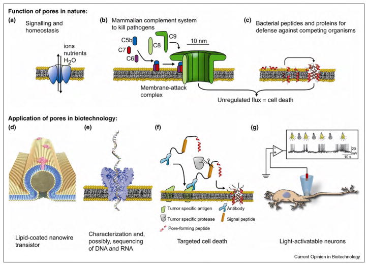
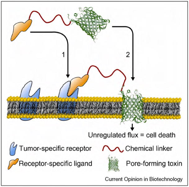
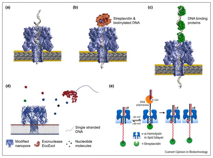
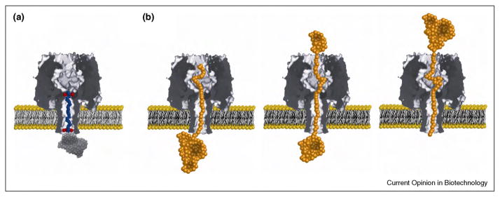
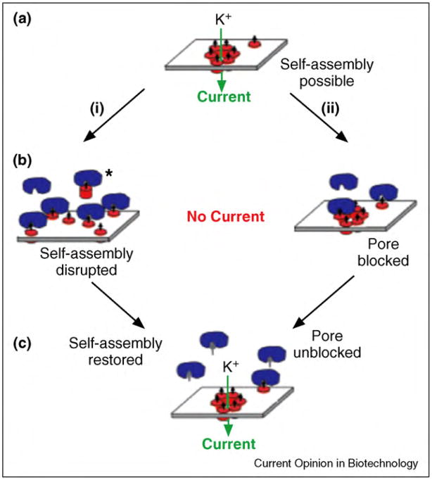
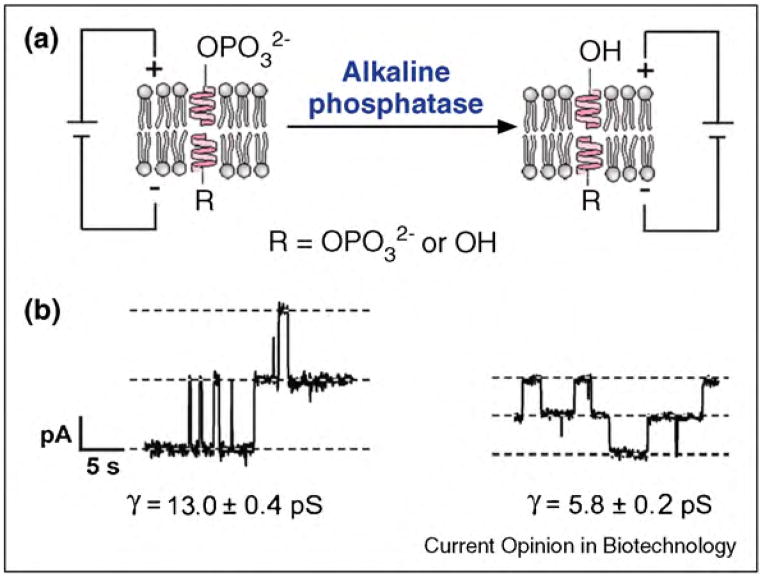
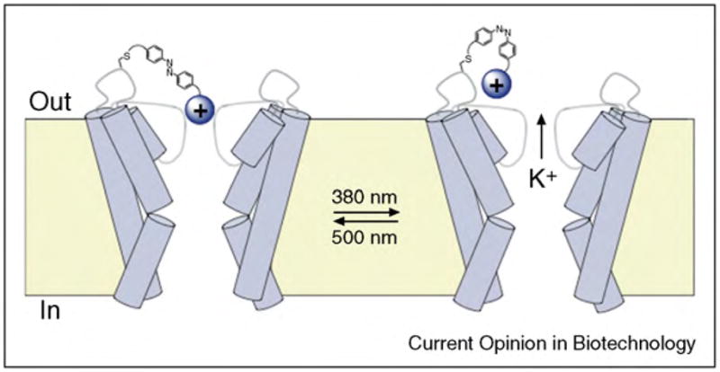
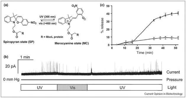
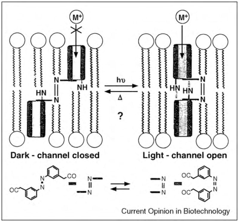
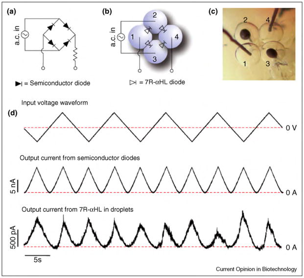
Biological protein pores and pore-forming peptides can generate a pathway for the flux of ions and other charged or polar molecules across cellular membranes. In nature, these nanopores have diverse and essential functions that range from maintaining cell homeostasis and participating in cell signaling to activating or killing cells. The combination of the nanoscale dimensions and sophisticated - often regulated - functionality of these biological pores make them particularly attractive for the growing field of nanobiotechnology. Applications range from single-molecule sensing to drug delivery and targeted killing of malignant cells. Potential future applications may include the use of nanopores for single strand DNA sequencing and for generating bio-inspired, and possibly, biocompatible visual detection systems and batteries. This article reviews the current state of applications of pore-forming peptides and proteins in nanomedicine, sensing, and nanoelectronics.


Figures

Figure 1
Figure 1
Functions of biological nanopores in nature and applications of these pores in nanobiotechnology. (a) Ion channel proteins transport ions across the plasma membrane of a cell for maintaining homeostasis in the cell and for signaling purposes. (b) Membrane-attack complexes form a lytic pore with a diameter of ~10 nm in the plasma membrane of a pathogen by self-assembly of the complement proteins C5b to C9. (c) Antibiotic peptides (here alamethicin [31]) insert into the membrane of target microbes and form lytic pores. (d) Bionanoelectronic device consisting of a silicon nanowire coated with a lipid bilayer with peptide pores. Image by Scott Dougherty, Lawrence Livermore National Laboratory. (e) Translocation of a single-stranded DNA molecule through an engineered bacterial porin, MspA, leads to partial blockage of the pore; translocation can be monitored by the resulting fluctuations in the ionic current through the pore. (f) Activation of multimeric pores by a tumor specific protease targets and kills malignant cells. (g) Remotely activated firing of neurons by engineered, light-activated ion channel proteins.
Figure 2
Figure 2
Cartoon illustrating a simple approach employed for targeted cytolysis of cancer cells that uses biological pores. A pore-forming peptide or protein is attached to a ligand that recognizes tumor specific receptor proteins. Once the ligand binds on the surface of a cancer cell, the pore-forming peptide or protein inserts into the membrane of the cell and forms a lytic pore.
Figure 3
Figure 3
Basic concept of using a multimeric pore, with a built-in ‘trigger’ system to target and kill cancer cells. Monomeric peptides are attached to monoclonal antibodies that recognize tumor specific antigens on targeted cancer cells. Upon this binding, tumor specific proteases secreted by these cancer cells recognize and cleave the peptide extensions on the monomers that inhibited their assembly to pores. The resulting active peptides can form cytolytic membrane pores and kill cancer cells.
Figure 4
Figure 4
Schematic drawing illustrating the principle of resistive-pulse sensing of analytes (green sphere) with α-hemolysin pores. The pores are engineered to contain an artificial binding site for the analyte in their lumen. In the presence of a transmembrane potential, binding of an analyte molecule results in a partial blockage of the pore; this modulation can be detected by the fluctuations in the ionic current passing through the pore. Figure reprinted from reference [3••] with permission.
Figure 5
Figure 5
Schematic illustration of the three main approaches to engineer α-hemolysin pores for sensing. (a) Genetic modification of the pore makes it possible to position desirable amino acid residues inside the lumen of the pore. (b) Placement of ring-shaped molecular adaptors such as cyclodextrins, inside its lumen. (c) Covalent attachment of a ligand-terminated PEG polymer into the lumen of the pore. Figure adapted from reference [3••] with permission.
Figure 6
Figure 6
Resistive-pulse sensing through protein pores (here α-hemolysin) makes it possible to determine the size of polymers in solution as well as to monitor the kinetics of polymer chain elongation. (a) Polymers of different molecular weight translocating through a protein pore result in transient current blockages of different magnitude. Figure adapted form reference [222] with permission. (b) Polymers that are linked covalently to the interior of a protein pore can be used to observe chemical reactions that lead to the addition of individual monomers; each added monomer decreases the current through the pore.
Figure 7
Figure 7
Overview of techniques that are being explored for sequencing single-stranded DNA or RNA with protein pores. (a) A segment of double-stranded DNA temporarily stops the translocation of long single-stranded DNA segments. This technique may be employed for de novo sequencing by hybridization [29,268]. (b) Streptavidin bound to single-stranded, biotinylated DNA can immobilize the DNA fragment in the pore, permitting a sufficiently long residence time for identification of individual base mutations [236,264,265,266]. (c) Proteins bound to DNA can slow the translocation of single-stranded DNA through pores facilitating identification of nuleotides [269]. (d) The pore α-hemolysin with a cyclodextrin adapter can be employed to distinguish between different nucleosides based on the magnitude of current blockages. In combination with an exonuclease to digest single-stranded DNA, this approach might allow sequencing [270••]. Figure adapted form reference [270••] with permission. (e) Single-stranded DNA that is attached to a biotinylated PEG-polymer on one side of the pore and to a complimentary DNA segment on the other side can be trapped within α-hemolysin pores. The activity of DNA polymerase adds individual nucleotides which increases the conductance of the pore because the PEG polymer chain, which has a smaller diameter than DNA, occupies more of the α-hemolysin pore after the addition of each nucleotide [271]. Figure adapted form reference [271] with permission.
Figure 8
Figure 8
Cartoon illustrating the concept of electrostatic traps to capture proteins in biological pores as well as the investigation of unfolding of a protein during translocation. (a) Negatively charged residues in the lumen of α-hemolysin pores can capture polypeptides that are positively charged. This effect can be used for selective capture of a large protein at the entrance of a pore [292]. (b) Cartoon illustrating the concept of unfolding of a protein at the entrance of a pore before translocation through the pore. Refolding of the protein on the other side of the pore may complete the process.
Figure 9
Figure 9
Concept of detection of protein–ligand-binding interactions using the antibiotic peptide gramicidin A. (a) A lipid bilayer tethered on a gold electrode contains tethered gramicidin monomers in one leaflet of the bilayer and free monomers with attached antigen-binding fragments (Fab) of antibodies in the other leaflet of the bilayer. In the absence of an analyte, dimerization of gramicidin monomers in the two leaflets leads to the formation of gramicidin pores across the bilayer and to an increase in the ionic conductivity of the membrane. Binding of analyte to the antibodies on the gramicidin monomers crosslinks these Fab molecules and limits the diffusion of bound gramicidin monomers within the outer leaflet of the bilayer. This interaction slows the formation of channel dimers and lowers the electrical conductivity of the membrane. Figure reprinted from reference [168••] with permission. (b) Binding of a protein (in this case avidin) to lipids with covalently attached ligands (in this case biotin lipids) in a planar lipid membrane results in a local distortion of the bilayer structure leading to detectable changes in the kinetics of formation of gramicidin pores (i.e. changes in lifetime and opening frequency). Figure reprinted from reference [195] with permission.
Figure 10
Figure 10
Basic concept of sensing protein–ligand interactions by disrupting the self-assembly of pore formers to a conducting pore. (a) Alamethicin monomers (red cylinders) with covalently attached ligands (small black arrows) self-assemble to form pores in a planar lipid bilayer as evident from single channel recordings. (b) Binding of a aprotein (here carbonic anhydrase II, shown in blue) to the ligand could have two consequences: (1) disruption of the pore, either by steric hindrance, or by removing the peptide from the bilayer, or (2) blockage of the mouth of the pore. In both cases, the binding interaction reduces the ionic current through the pore. (c) Addition of competitive ligand (small gray arrows) to the solution leads to binding of free ligand to the proteins and to the release of alamethicin peptides, which lead to pore formation. Figure reprinted from reference [24] with permission.
Figure 11
Figure 11
Concept of a gramicidin-based sensor for monitoring the enzymatic activity of alkaline phosphatase in situ. (a) Enzyme-catalyzed hydrolysis of a negatively charged phosphate group from gramicidin-phosphate to a gramicidin-derivative with a neutral alcohol group. (b) Corresponding current versus time recordings. At low ionic strength in the recording buffer, the single channel conductance, γ, through pores of the neutral gramicidin derivative is significantly smaller than the conductance through pores of the charged gramicidin-phosphate. This effect results from electrostatic accumulation of monovalent cations (which carry the charge through gramicidin pores) near the pore entrance of the negatively charged gramicidin-phosphate. Figure reprinted from reference [22] with permission.
Figure 12
Figure 12
Schematic illustration of an α-hemolysin-based platform for monitoring the cleavage of a peptide by a protease. Before addition of the protease, only substrate molecules (in this case, residues 10–20 of amyloid-β peptides) pass through the pore and produce characteristic blockage events as shown in pathway (a). Addition of the protease to the solution results in cleavage of the substrate peptides, producing smaller peptide fragments. Passage of the resulting fragments through the engineered α-hemolysin pore can be detected through blockage events that are significantly different in amplitude and length from those produced by the substrate peptide, as shown in pathway (b). Figure reprinted from reference [112] with permission.
Figure 13
Figure 13
Basic concept of monitoring the activity of a membrane-active enzyme, phospholipase D (PLD), on planar lipid bilayers. Enzyme activity is recorded by changes in single channel conductance of gramicidin pores. (a) As PLD hydrolyzes electrically neutral phosphatidylcholine (PC) lipids and produces negatively charged phosphatidic acid (PA) lipids, the electrostatic accumulation of cations close to the membrane surface leads to a significant increase in channel conductance of gramicidin pores. Negative charges are shown in red, and positive ions are shown in blue. (b) Corresponding current versus time recordings before and after addition of PLD. Figure adapted from reference [23] with permission.
Figure 14
Figure 14
Monitoring chemical reactions on functional groups attached to gramicidin peptides through single channel recordings. (a) Illustration of the stepwise conversion of gramicidin carrying a Boc-protected glycine group (left) to gramicidin carrying a glycolic acid group (right) in the presence of different reagents. (b) Corresponding single channel recordings with characteristic conductance values of each derivative. Figure reprinted from reference [33] with permission.
Figure 15
Figure 15
Hydrophobic mismatch between a membrane and an embedded transmembrane protein or peptide. (a) Cartoon of a protein that has a transmembrane segment of length (h) that is shorter than the distance (d) across the hydrophobic core of the lipid bilayer, and the resulting compression of the bilayer. (b) The transmembrane segment of the protein has the same length as the hydrophobic core of the lipid bilayer. (c) The hydrophobic segment of the protein is longer than the membrane can accommodate without generating the energetic expense associated with stretching and bending forces.
Figure 16
Figure 16
Cartoon illustrating the concept of remote control of neuronal firing by light. Expression of a light-activated ion channel (in this case, a modified potassium channel) in rat hippocampal neurons made these neurons sensitive to light as confirmed by current clamp recordings. Exposure to light with a wavelength of 500 nm resulted in spontaneous action potentials, while exposure to light with a wavelength of 380 nm silenced these action potentials. The inset shows a current versus time trace as adapted from reference [34••] with permission.
Fig. 17
Fig. 17
Example of a light-gated potassium ion channel generated by covalent attachment of a pore-blocking group via a photoisomerizable linker to the exit of the channel protein. A covalently linked, positively charged group moved either close to, or away from, the exit of the channel depending on the light-induced configuration of the linker. Figure adapted from reference [34••] with permission.
Figure 18
Figure 18
Example of a photo-gated, MscL ion channel generated by incorporation of a photo-activatable spiropyran group into the lumen of the pore. (a) Chemical structure and reversible photo-induced conversion of a non-polar spiropyran molecule to a polar merocyanine conformational state. (b) Current versus time trace of a modified MscL channel carrying a covalently attached spiropyran group inside its lumen. The frequency of channel openings decreased significantly upon exposure of these channels to visible light compared to exposure to ultraviolet light. (c) Results of a leakage assay demonstrating the release profile of fluorescent calcein molecules from proteoliposomes that contained modified MscL pores when exposed to (■) ultraviolet, or (□) visible light. Figure adapted from reference [194••] with permission.
Figure 19
Figure 19
Concept of a light-gated gramicidin channel with a diazobenzene linker that was used to control the alignment of two gramicidin monomers within a membrane. Figure reprinted from reference [336] with permission.
Figure 20
Figure 20
Current rectification in man-made electronic circuits, in biology and nanobiotechnology. (a) Semiconductor diode with a p–n junction that allows electrons to flow only in one direction. (b) Inward-rectifying potassium channel (Kir2.2) that conducts potassium ions most efficiently in one direction. The current versus voltage graph is from reference [48] with permission. (c) Two different derivatives of gramicidin (one carrying a positive charge at its C-terminus and the other carrying a negative charge at its C-terminus) form a pore across a membrane. This heterodimeric gramicidin pore acts as the smallest nanofluidic diode and rectifies current. The current versus voltage graph is from reference [350] with permission.
Figure 21
Figure 21
Comparison between the performance of a full-wave bridge rectifier based on semiconductor diodes and a full-wave rectifier based on engineered biological pores. (a) Circuit diagram of a bridge rectifier with four diodes to achieve full-wave rectification. (b) Illustration of a four-droplet network to form a bridge rectifier with the mutant α-hemolysin protein 7R-αHL. (c) Photograph of the system in (b). (d) Electric properties of the droplet network circuit: input 0.1 Hz triangular wave (top); output current observed from a bridge rectifier using semiconductor diodes (middle); output current from a bridge rectifier system based on a droplet interface bilayer network (bottom). Figure reprinted from reference [109] with permission.
Figure 22
Figure 22
Design and function of bilayer-coated nanowires. (a) Schematic illustration of bilayer-coated nanowires that are connected to microfabricated electrodes, which constitute the source (S) and drain (D). The insets of this figure show a cartoon of the bilayer membranes with embedded ion channel-forming peptides. (b) Graph illustrating the change of conductance of bilayer-coated nanowires as the pH is altered from 5 to 7. Red curve: nanowire device without a lipid bilayer coating. Blue curve: nanowire device with a lipid bilayer coating and incorporated gramicidin pores. Black curve: nanowire device with a lipid bilayer coating and incorporated gramicidin pores in the presence of channel-blocking calcium ions. Figure adapted from reference [28] with permission.
Figure 23
Figure 23
Block diagram of a droplet interface bilayer network with feedback current control. Figure adapted from reference [357] with permission.
Figure 24
Figure 24
Schematic illustration of an experimental setup used to form a bio-inspired battery and current versus time trace recorded from this setup. Arrows indicate removal of red droplets one at a time. Figure adapted from reference [26••] with permission.
Figure 25
Figure 25
Diagram of a droplet interface bilayer network that can sense light and current versus time trace showing a large upward spike when laser was switched on (green upward arrows) followed by a rapid decay to a steady-state current of ~5 pA. When the laser was turned off (black downward arrows), the current temporarily reached negative values before returning to baseline. Figure adapted from reference [26••] with permission.

References

    1. Aidley DJ, Stanfield PR. Ion Channels: Molecules in Action. Cambridge: Cambridge University Press; 1996.
    1. Alberts B, Johnson A, Lewis J, Raff M, Roberts K, Walter P. Molecular Biology of the Cell. 4. New York: Garland Science; 2002.
    1. Bayley H, Cremer PS. Stochastic sensors inspired by biology. Nature. 2001;413:226–230. This article provides an insight to the application of engineered biological pores for development of rapid and sensitive biosensors with a focus on the application of these pores in stochastic sensing. - PubMed
    1. Panchal RG, Smart ML, Bowser DN, Williams DA, Petrou S. Pore-forming proteins and their application in biotechnology. Curr Pharm Biotechnol. 2002;3:99–115. This excellent review introduces a number of channel proteins and pore-forming peptides that have been used in biotechnology. It also provides a brief overview of the applications of these pores in medicine and sensing. - PubMed
    1. Estes DJ, Memarsadeghi S, Lundy SK, Marti F, Mikol DD, Fox DA, Mayer M. High-throughput profiling of ion channel activity in primary human lymphocytes. Anal Chem. 2008;80:3728–3735. - PubMed

Publication types