

Mitral annulus segmentation from 3D ultrasound using graph cuts
- PMID: 20562042
- PMCID: PMC3122108
- DOI: 10.1109/TMI.2010.2050595
Mitral annulus segmentation from 3D ultrasound using graph cuts
Abstract
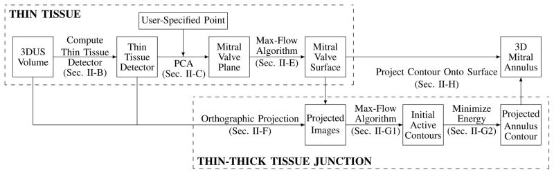
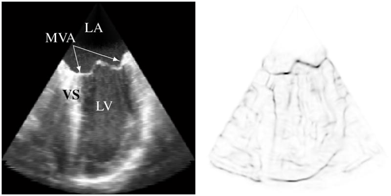
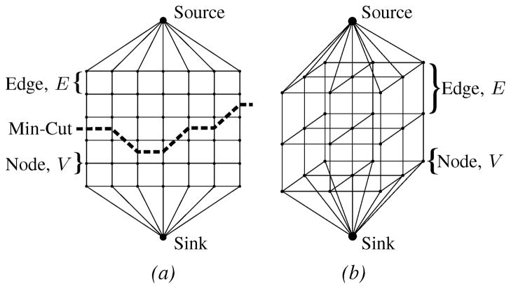
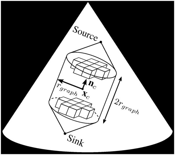
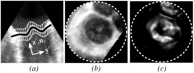
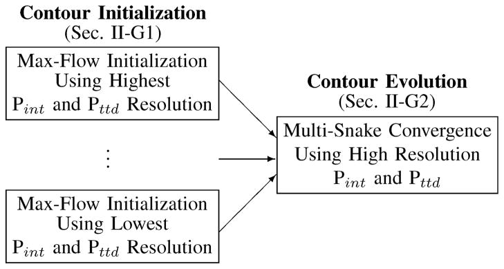
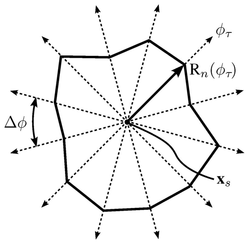
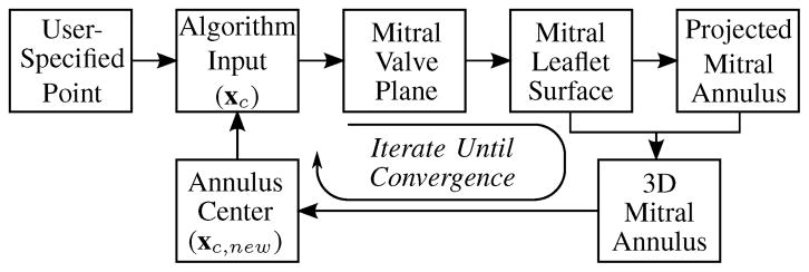
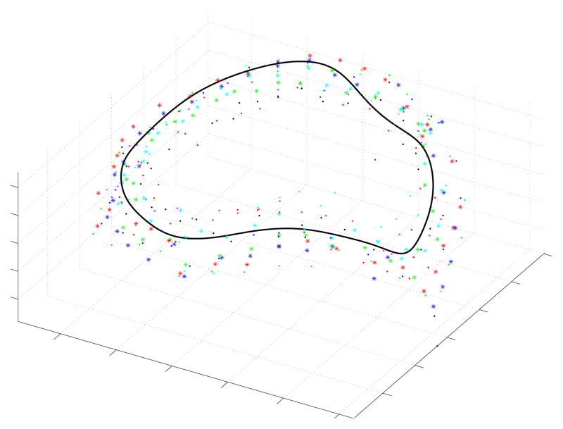
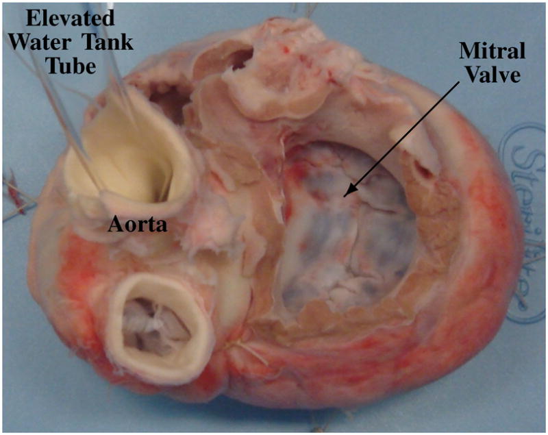
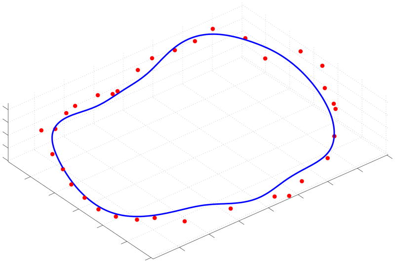
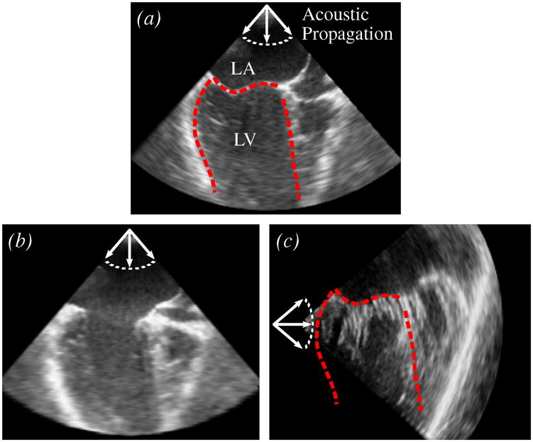
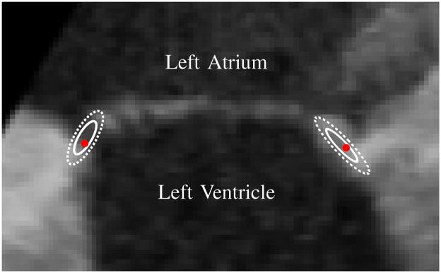
The shape of the mitral valve annulus is used in diagnostic and modeling applications, yet methods to accurately and reproducibly delineate the annulus are limited. This paper presents a mitral annulus segmentation algorithm designed for closed mitral valves which locates the annulus in three-dimensional ultrasound using only a single user-specified point near the center of the valve. The algorithm first constructs a surface at the location of the thin leaflets, and then locates the annulus by finding where the thin leaflet tissue meets the thicker heart wall. The algorithm iterates until convergence metrics are satisfied, resulting in an operator-independent mitral annulus segmentation. The accuracy of the algorithm was assessed from both a diagnostic and surgical standpoint by comparing the algorithm's results to delineations made by a group of experts on clinical ultrasound images of the mitral valve, and to delineations made by an expert with a surgical view of the mitral annulus on excised porcine hearts using an electromagnetically tracked pointer. In the former study, the algorithm was statistically indistinguishable from the best performing expert (p=0.85) and had an average RMS difference of 1.81+/-0.78 mm to the expert average. In the latter, the average RMS difference between the algorithm's annulus and the electromagnetically tracked points across six hearts was 1.19+/-0.17 mm .
Figures

References
-
- Levine R, Handschumacher M, Sanfilippo A, Hagege A, Harrigan P, Marshall J, et al. Three-dimensional echocardiographic reconstruction of the mitral valve, with implications for the diagnosis of mitral valve prolapse. Circulation. 1989;80(3):589–598. - PubMed
-
- Flachskampf F, Chandra S, Gaddipatti A, Levine R, Weyman A, Ameling W, et al. Analysis of shape and motion of the mitral annulus in subjects with and without cardiomyopathy by echocardiographic 3-dimensional reconstruction. Journal of the American Society of Echocardiography. 2000;13(4):277–287. - PubMed
-
- Kaplan S, Bashein G, Sheehan F, Legget M, Munt B, Li X, et al. Three-dimensional echocardiographic assessment of annular shape changes in the normal and regurgitant mitral valve. American Heart Journal. 2000;139(3):378–387. - PubMed
-
- Kaji S, Nasu M, Yamamuro A, Tanabe K, Nagai K, Tani T, et al. Annular Geometry in Patients With Chronic Ischemic Mitral Regurgitation: Three-Dimensional Magnetic Resonance Imaging Study. Circulation. 2005;112(9 Suppl):I409–I414. - PubMed
-
- Popovic Z, Martin M, Fukamachi K, Inoue M, Kwan J, Doi K, et al. Mitral annulus size links ventricular dilatation to functional mitral regurgitation. Journal of the American Society of Echocardiography. 2005;18(9):959–963. - PubMed
Publication types
MeSH terms
Grants and funding
LinkOut - more resources
Full Text Sources
Other Literature Sources
