. 2010 Aug 27;285(35):27429-27439.
doi: 10.1074/jbc.M110.142752. Epub 2010 Jun 18.

Inhibition of STAT3 signaling blocks the anti-apoptotic activity of IL-6 in human liver cancer cells

Affiliations

Inhibition of STAT3 signaling blocks the anti-apoptotic activity of IL-6 in human liver cancer cells

Yan Liu et al. J Biol Chem. .

Abstract

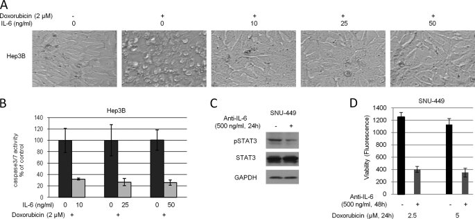
Interleukin-6 (IL-6) is a multifunctional cytokine, which may block apoptosis during inflammation to protect cells under very toxic conditions. However, IL-6 also activates STAT3 in many types of human cancer. Recent studies demonstrate that high levels of IL-6 are associated with hepatocellular carcinoma, the most common type of liver cancer. Here we reported that IL-6 promoted survival of human liver cancer cells through activating STAT3 in response to doxorubicin treatment. Endogenous IL-6 levels in SNU-449 cells were higher than in Hep3B cells. Meanwhile, SNU-449 cells were more resistant to doxorubicin than Hep3B cells. Addition of IL-6 induced STAT3 activation in Hep3B cells and led to protection against doxorubicin. In contrast, neutralizing IL-6 with anti-IL-6 antibody decreased survival of SNU-449 cells in response to doxorubicin. To elucidate the mechanism of the anti-apoptotic function of IL-6, we investigated if STAT3 mediated this drug resistance. Targeting STAT3 with STAT3 siRNA reduced the protection of IL-6 against doxorubicin-induced apoptosis, indicating that STAT3 signaling contributed to the anti-apoptotic effect of IL-6. Moreover, we further explored if a STAT3 small molecule inhibitor could abolish this anti-apoptotic effect. LLL12, a STAT3 small molecule inhibitor, blocked IL-6-induced STAT3 phosphorylation, resulting in attenuation of the anti-apoptotic activity of IL-6. Finally, neutralization of endogenous IL-6 with anti-IL-6 antibody or blockade of STAT3 with LLL12 lowered the recovery in SNU-449 cells after doxorubicin treatment. Therefore, our results demonstrated that targeting STAT3 signaling could interrupt the anti-apoptotic function of IL-6 in human liver cancer cells.


Figures

FIGURE 1.
FIGURE 1.
IL-6 induces STAT3 phosphorylation. A, human hepatocyte, Hep3B, SNU-387, SNU-398, and SNU-449 cells were cultured in Hepatocyte Medium for 24 h. Endogenous IL-6 was accessed by IL-6 ELISA assay. B, Hep3B and SNU-398 cells were cultured in serum-free medium. After 24 h of serum deprivation, cells were treated with various concentrations of IL-6 for 30 min. STAT3 phosphorylation and total STAT3 were detected by Western blot. C, Hep3B and SNU-398 cells were cultured under the same conditions and then were treated with 50 ng/ml of IL-6 for 30 min. The distribution of phosphorylated STAT3 was analyzed by immunofluorescence.
FIGURE 2.
FIGURE 2.
SNU-449 cells are more doxorubicin-resistant than Hep3B cells. A, SNU-449 cells and Hep3B cells were treated with various concentrations of doxorubicin. After 24 h, morphological examination was performed. B, SNU-449 cells and Hep3B cells were treated under the same conditions. After 48 h, cell viability assay was performed. The data showed the percent decrease in cell viability compared with untreated cells and represented three independent results. C, endogenous phosphorylated STAT3 and total STAT3 in Hep3B and SNU-449 cells were analyzed by Western blot.
FIGURE 3.
FIGURE 3.
IL-6 protects cells against doxorubicin-induced apoptosis. A, Hep3B cells were pretreated with 2 μm doxorubicin for 2 h followed by various concentrations of IL-6 for 6 h. Morphological examination was performed after treatment. B, Hep3B cells were treated under the same conditions. After 24 h, caspase3/7 activity was analyzed. The data showed the percent decrease in csapase3/7 activity compared with control cells and represented three independent results. C, SNU-449 cells were treated with 500 ng/ml of anti-IL-6 antibody or control IgG for 24 h. After the incubation, whole cell lysates were extracted. Phosphorylated STAT3 and GAPDH were detected by Western blot. D, SNU-449 cells were pretreated with 500 ng/ml of anti-IL-6 antibody or control IgG for 24 h. After the pretreatment, cells were further treated with doxorubicin for 24 h. After the 24 h treatment, cell viability was measured. The data represented eight independent results.
FIGURE 4.
FIGURE 4.
Inhibition of phosphorylated STAT3 blocks IL-6-induced cell survival. A, two siRNAs targeting STAT3 were transfected into Hep3B cells. After overnight incubation, the cells were cultured in serum-free medium for 24 h followed by IL-6 for 30 min. Phosphorylated STAT3, phosphorylated AKT, and GAPDH were detected by Western blot. B, STAT3 siRNA-transfected Hep3B cells were treated with 2 μm doxorubicin for 2 h followed by 50 ng/ml of IL-6 for 6 h. Morphological examination was performed after treatment. C, STAT3 siRNA-transfected Hep3B cells were pretreated with doxorubicin for 2 h followed by 50 ng/ml of IL-6 for 24 h. Caspase3/7 activity was assayed. The data showed the percent increase in caspase3/7 compared with scrambled siRNA-transfected control groups and represented three independent experiments. *, p < 0.02.
FIGURE 5.
FIGURE 5.
A STAT3 small molecule inhibitor LLL12 blocks IL-6-induced STAT3 phosphorylation. A, Hep3B and SNU-398 cells were cultured in serum-free medium for 24 h and then were pretreated with DMSO or LLL12 for 2 h followed by IL-6 for 30 min. Phosphorylated STAT3 and total STAT3 were detected by Western blot. B, Hep3B and SNU-398 cells were cultured in serum-free medium for 24 h and then were pretreated with LLL12 for 2 h followed by IFN-γ for 30 min. The expression levels of phosphorylated STAT1, and total STAT1 were analyzed by Western blot. C and D, Hep3B cells were treated under the same conditions, and the distribution of phosphorylated STAT3 and total STAT3 was analyzed by immunofluorescence.
FIGURE 6.
FIGURE 6.
The STAT3 small molecule inhibitor LLL12 blocks STAT3 phosphorylation in a dose- and time-dependent manner. A, Hep3B and SNU-398 cells were cultured in serum-free medium for 24 h and then were pretreated with various concentrations of LLL12 (0.5–5 μm) for 2 h followed by IL-6 for 30 min. Phosphorylated STAT3 and total STAT3 were detected by Western blot. B, Hep3B and SNU-398 cells were cultured under the same conditions and then were treated with 5 μm LLL12 for different time points (0–2 h). After pretreatment, IL-6 was added to the cultured cells. Phosphorylated STAT3 and total STAT3 were detected by Western blot. C, Hep3B cells were cultured in serum-free medium for 24 h and then were pretreated with 5 μm LLL12 for 2 h. After the 2 h pretreatment, medium was discarded, and fresh medium without LLL12 was added. After the indicated incubation (0–24 h), the cells were treated with IL-6 for 30 min. Phosphorylated STAT3 and total STAT3 were detected by Western blot. D, Hep3B cells were cultured in serum-free medium for 24 h and then were pretreated with 5 μm LLL12 for 2 h. After 2 h of pretreatment, the medium was discarded, and fresh medium without LLL12 was added. Cycloheximide was also added into the medium to block protein synthesis. After 12 h, the cells were treated with IL-6 for 30 min. Phosphorylated STAT3 and total STAT3 were detected by Western blot.
FIGURE 7.
FIGURE 7.
LLL12 decreases IL-6-induced cell survival. A, Hep3B cells were pretreated with 0.5 μm LLL12 for 2 h. After the pretreatment, LLL12 was removed, and fresh medium was added. The cells were treated as indicated. After overnight treatment, morphological examination was performed. B, results from A were quantified by cell viability assay. C and D, Hep3B and SNU-398 cells were pretreated with 0.5 μm LLL12 for 2 h. After the pretreatment, cells were treated with IL-6 for 30 min. The levels of JAK1, JAK2, GP130, p-AKT, and p-ERK1/2 were analyzed by Western blot.
FIGURE 8.
FIGURE 8.
Blockade of IL-6 or STAT3 reduces cell recovery from doxorubicin. SNU-449 cells were treated with 2 μm doxorubicin, anti-IL-6 antibody, or LLL12 as indicated. After overnight treatment, live cells were counted and 5,000 cells were re-plated. Cells were treated with control IgG, anti-IL-6 antibody, or LLL12, respectively. After 7 days, cells were fixed and stained with 1% crystal violet. A, pictures were captured under microscopy at different magnification. B, original plates were scanned. Three independent experiments were performed.
FIGURE 9.
FIGURE 9.
Blockade of IL-6 or STAT3 does not affect cell viability in human primary hepatocytes treated with doxorubicin. A, human primary hepatocytes were treated with 500 ng/ml of anti-IL-6 antibody or 0.5 μm LLL12 for 2 h and 24 h, respectively. After 24 h, cell viability was measured. Non-treated cells (NT) were used as control. The data represented three independent results. B, human primary hepatocytes were pretreated with 500 ng/ml of anti-IL-6 antibody or 0.5 μm LLL12 for 2 h. After the pretreatment, LLL12 was discarded and fresh medium with different concentrations of doxorubicin was added. After 24 h, cell viability was measured. The data represented three independent results.

Similar articles

Cited by

References

    1. Hodge D. R., Hurt E. M., Farrar W. L. (2005) Eur. J. Cancer 41, 2502–2512 - PubMed
    1. Grivennikov S., Karin E., Terzic J., Mucida D., Yu G. Y., Vallabhapurapu S., Scheller J., Rose-John S., Cheroutre H., Eckmann L., Karin M. (2009) Cancer Cell 15, 103–113 - PMC - PubMed
    1. Schafer Z. T., Brugge J. S. (2007) J. Clin. Invest. 117, 3660–3663 - PMC - PubMed
    1. Bharti A. C., Donato N., Aggarwal B. B. (2003) J. Immunol. 171, 3863–3871 - PubMed
    1. Sansone P., Storci G., Tavolari S., Guarnieri T., Giovannini C., Taffurelli M., Ceccarelli C., Santini D., Paterini P., Marcu K. B., Chieco P., Bonafè M. (2007) J. Clin. Invest. 117, 3988–4002 - PMC - PubMed

Publication types

MeSH terms