. 2010 Aug 27;285(35):27067-27077.
doi: 10.1074/jbc.M110.110072. Epub 2010 Jun 18.

Allosteric block of KCa2 channels by apamin

Affiliations

Allosteric block of KCa2 channels by apamin

Cédric Lamy et al. J Biol Chem. .

Abstract

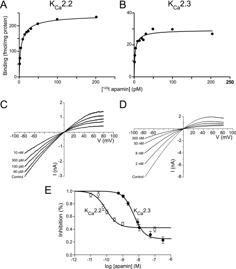
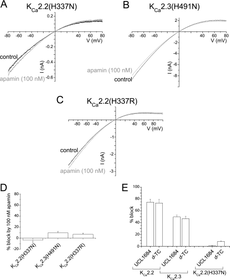
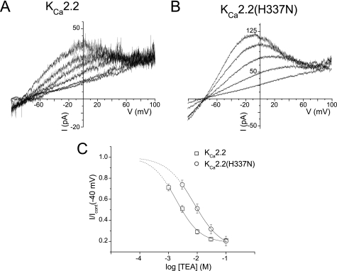
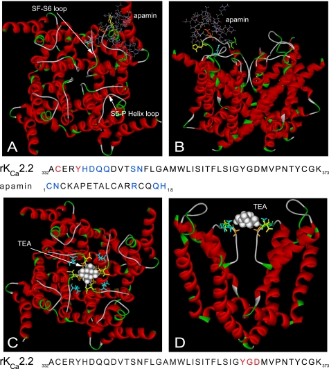
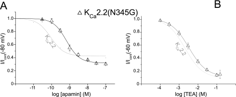
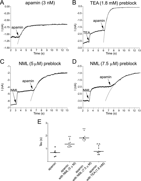
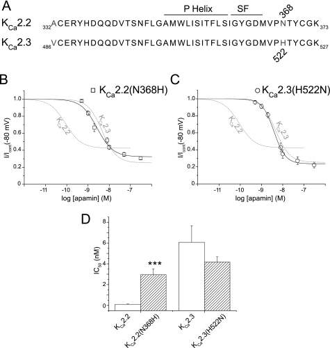
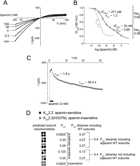
Activation of small conductance calcium-activated potassium (K(Ca)2) channels can regulate neuronal firing and synaptic plasticity. They are characterized by their high sensitivity to the bee venom toxin apamin, but the mechanism of block is not understood. For example, apamin binds to both K(Ca)2.2 and K(Ca)2.3 with the same high affinity (K(D) approximately 5 pM for both subtypes) but requires significantly higher concentrations to block functional current (IC(50) values of approximately 100 pM and approximately 5 nM, respectively). This suggests that steps beyond binding are needed for channel block to occur. We have combined patch clamp and binding experiments on cell lines with molecular modeling and mutagenesis to gain more insight into the mechanism of action of the toxin. An outer pore histidine residue common to both subtypes was found to be critical for both binding and block by the toxin but not for block by tetraethylammonium (TEA) ions. These data indicated that apamin blocks K(Ca)2 channels by binding to a site distinct from that used by TEA, supported by a finding that the onset of block by apamin was not affected by the presence of TEA. Structural modeling of ligand-channel interaction indicated that TEA binds deep within the channel pore, which contrasted with apamin being modeled to interact with the channel outer pore by utilizing the outer pore histidine residue. This multidisciplinary approach suggested that apamin does not behave as a classical pore blocker but blocks using an allosteric mechanism that is consistent with observed differences between binding affinity and potency of block.


Figures

FIGURE 1.
FIGURE 1.
Binding and block of KCa2.2 and KCa2.3 channel current by apamin. A and B, representative examples of saturation relationships for 125I-apamin binding to expressed KCa2.2 (A) or KCa2.3 (B) subunits. C and D, whole-cell macroscopic currents derived from ramps from −80 to 80 mV imposed on voltage-clamped HEK293 cells expressing KCa2.2 (C) and (D) KCa2.3 subunits. The application of increasing concentrations of apamin inhibited macroscopic current. E, concentration-inhibition relationships for apamin inhibition of expressed KCa2.2 and KCa2.3 current.
FIGURE 2.
FIGURE 2.
His-337/His-491 residues are critical for block of KCa2.2 and KCa2.3 currents by apamin and organic blockers. A–C, outside-out patch (A and C) and whole-cell (B) macroscopic currents evoked by ramps from −80 to 80 mV in the absence (black trace) and the presence of a supramaximal concentration of apamin (100 nm) (gray trace). Mutation of the outer pore His residue in KCa2.2(H337N) (A) and KCa2.3(H491N) (B) produced currents that were insensitive to the bee venom toxin. Mutation of His-337 in KCa2.2 to the positively charged arginine (H337R) also produced currents that were apamin-insensitive (C). D, graph showing the mean ± S.E. inhibition by 100 nm apamin of macroscopic current from each mutant. E, graph showing the lack of block of KCa2.2(H337N) by the organic blockers UCL1684 (20 nm) and d-TC (100 μm) when compared with block of WT KCa2.2- and KCa2.3-mediated current.
FIGURE 3.
FIGURE 3.
Block of KCa2.2 and KCa2.2(H337N) channels by extracellular TEA. A and B, representative traces from outside-out patches of expressed KCa2.2 (A) and KCa2.2(H337N) (B) channel currents evoked by voltage ramps from −100 to 100 mV in the absence and presence of increasing concentrations of extracellular TEA. Experiments were performed in low (5 mm) extracellular [K+], with increasing concentrations of TEA being substituted for NaCl. C, concentration-inhibition relationships for block of wild-type KCa2.2 and mutant KCa2.2(H337N) current by TEA (see “Results” for details).
FIGURE 4.
FIGURE 4.
Structural modeling of the interaction between the KCa2.2 channel and apamin or TEA. A and B, top down (A) and side (B)views of apamin docked to the outer pore region of KCa2.2. Channel residues discussed under “Results” are highlighted as follows: His-337 (yellow), Gln-339 (orange), Asn-345 (green), and Asn-368 (cyan). Residues within the channel outer pore and apamin that make contact by hydrogen bonds are colored blue, whereas those channel residues making electrostatic interactions are in red. C and D, top down (C) and side (D) views of the interaction between TEA and KCa2.2. The quaternary ammonium ion is modeled to interact within the inner pore of the channel by electrostatic interactions with the C=O group of Tyr-362 (distance between C=O and N+ ∼4 Å), and the ethyl groups of TEA interact via van der Waals contacts with Gly-363 (orange), Asp-364 (yellow), and Val-366 (cyan).
FIGURE 5.
FIGURE 5.
Reduced sensitivity to block by apamin displayed by KCa2.2(N345G). A, concentration-inhibition relationship for block of expressed KCa2.2(N345G) channel current by apamin. The dashed gray curve shows the sensitivity of block of wild-type KCa2.2 current for comparison. B, concentration-inhibition relationship for block of KCa2.2(N345G) current by extracellular TEA. The relationship of block by TEA of wild-type KCa2.2 current is shown for comparison (dashed gray line), showing that the channel mutation had little effect upon block by the quaternary ion.
FIGURE 6.
FIGURE 6.
Apamin and TEA block KCa2.3 current by using non-interacting binding sites. A, example trace of whole-cell holding current recorded at −80 mV from a cell expressing KCa2.3 channels and bathed in high extracellular K+ solution. Fast application of apamin (3 nm) produced a block with a τon of 0.70 ± 0.12 s. Fits are shown as gray lines in all panels. B, membrane current recorded at −50 mV from a cell expressing KCa2.3 channel subunits. Fast application of TEA (1.8 mm) blocked ∼30% of current but had no effect on the time course of subsequent block by fast applied apamin (3 nm)(τon of 0.79 ± 0.08 s). C, fast application of NML (5 μm) blocked ∼40% of expressed KCa2.3 current, with the presence of this channel blocker slowing the rate of block by the subsequent fast application of apamin (τon slowed to 1.36 ± 0.08 s). D, a greater slowing of the rate of block by apamin was produced by a higher concentration of NML, with τon of block by apamin (3 nm) being slowed to 1.84 ± 0.09 s in 7.5 μm NML. E, graph showing individual values from all experiments of the τon (Tau) of block by apamin (3 nm) applied in the absence or presence of either TEA (1.8 mm) or NML (5 and 7.5 μm). A one-way analysis of variance showed that τon values were significantly different from each other (F = 37, p < 0.001). Tukey's post-hoc tests showed that the τon of apamin was unaffected by TEA (p > 0.05), but was significantly affected by both concentrations of NML (p < 0.05). n. s., not significant. ***, significant with p < 0.001.
FIGURE 7.
FIGURE 7.
Pore-mimicking mutants reveal differences between KCa2.2 and KCa2.3 structure. A, sequence alignment of the pore regions of KCa2.2 and KCa2.3 channels, with residues that differ highlighted in gray. SF, channel selectivity filter. B, concentration-inhibition relationships of KCa2.2(N368H), a mutation that produced a KCa2.2 channel whose pore mimicked KCa2.3. This mutation produced a channel current that was blocked by apamin with a sensitivity that was similar to that seen with wild-type KCa2.3 current. The relationships of block by apamin of wild-type KCa2.2 and KCa2.3 currents are shown for comparison in dashed gray. C, mutation of the outer pore of KCa2.3 to mimic KCa2.2 (KCa2.3(H522N)) produced a current that was blocked by apamin with a sensitivity that was not significantly different from wild-type KCa2.3 current. The relationships of block by apamin of wild-type KCa2.2 and KCa2.3 currents are shown for comparison in dashed gray. D, bar chart displaying the IC50 values showing the reduction in sensitivity in KCa2.2(N368H) when compared with WT KCa2.2(p < 0.0001, n = 7–10). No significant shift was observed for KCa2.3(H522N) when compared with WT KCa2.3 (p > 0.05, n = 8–12).
FIGURE 8.
FIGURE 8.
Co-expression of apamin-sensitive wild-type KCa2.2 and apamin-insensitive KCa2.2(H337N) produced heteromeric channels that displayed distinct sensitivities to apamin. A, representative outside-out patch current traces evoked by voltage ramps from −100 to 100 mV, in the absence (con, control) and presence of increasing concentrations of apamin. B, concentration-inhibition relationship for block of current produced by co-expression of wild-type KCa2.2 and mutant KCa2.2(H337N) channels by apamin. Mean data were fit with a two component Hill equation, with IC50 values of ∼270 pm and 33 nm, demonstrating that heteromeric channels were expressed. frac, fraction. C, example trace of outside-out patch holding current (Vh −80 mV) from a cell co-expressing wild-type and mutant KCa2.2 channels during the rapid application and removal of apamin (3 nm). Current was blocked by apamin with an exponential time course (not shown), with the recovery from block being best described by the sum of two exponential components of taus: τoff,1, 1.8 s, and τoff,2, 36.4 s. These data suggest that apamin is interacting with two populations of channels that possess different sensitivities to block by the toxin. D, the probabilities of occurrence (Pocc) of different predicted stoichiometries of channel subunit assembly were calculated assuming that the probability of the inclusion of KCa2.2 and KCa2.2(H337N) subunits into the channel tetramer was equivalent.

Similar articles

Cited by

References

    1. Blatz A. L., Magleby K. L. (1986) Nature 323, 718–720 - PubMed
    1. Vincent J. P., Schweitz H., Lazdunski M. (1975) Biochemistry 14, 2521–2525 - PubMed
    1. Pease J. H., Wemmer D. E. (1988) Biochemistry 27, 8491–8498 - PubMed
    1. Labbé-Jullié C., Granier C., Albericio F., Defendini M. L., Ceard B., Rochat H., Van Rietschoten J. (1991) Eur. J. Biochem. 196, 639–645 - PubMed
    1. Köhler M., Hirschberg B., Bond C. T., Kinzie J. M., Marrion N. V., Maylie J., Adelman J. P. (1996) Science 273, 1709–1714 - PubMed

Publication types

MeSH terms

Substances