. 2010 Oct;95(10):1745-53.
doi: 10.3324/haematol.2009.020958. Epub 2010 Jun 18.

Platelet-derived growth factor enhances platelet recovery in a murine model of radiation-induced thrombocytopenia and reduces apoptosis in megakaryocytes via its receptors and the PI3-k/Akt pathway

Affiliations

Platelet-derived growth factor enhances platelet recovery in a murine model of radiation-induced thrombocytopenia and reduces apoptosis in megakaryocytes via its receptors and the PI3-k/Akt pathway

Jie Yu Ye et al. Haematologica. 2010 Oct.

Abstract

Background: Platelet-derived growth factor is involved in the regulation of hematopoiesis. Imatinib mesylate, a platelet-derived growth factor receptor inhibitor, induces thrombocytopenia in a significant proportion of patients with chronic myeloid leukemia. Although our previous studies showed that platelet-derived growth factor enhances megakaryocytopoiesis in vitro, the in vivo effect of platelet-derived growth factor in a model of radiation-induced thrombocytopenia has not been reported.

Design and methods: In this study, we investigated the effect of platelet-derived growth factor on hematopoietic stem/progenitor cells and platelet production using an irradiated-mouse model. We also explored the potential molecular mechanisms of platelet-derived growth factor on thrombopoiesis in M-07e cells.

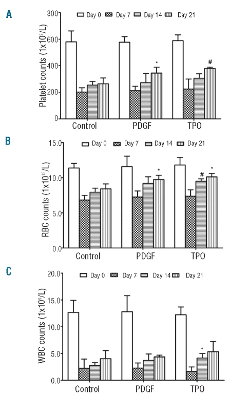
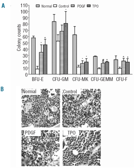
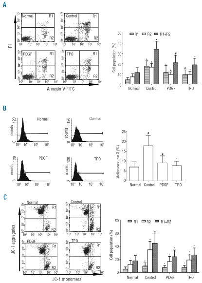
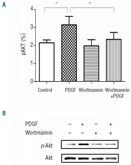
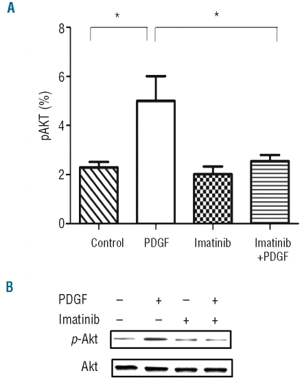
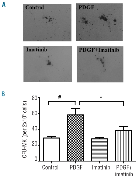
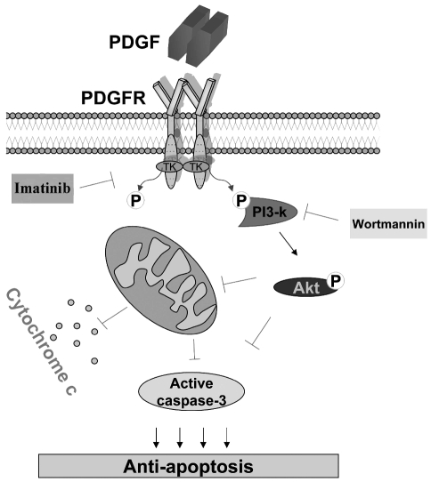
Results: Platelet-derived growth factor, like thrombopoietin, significantly promoted the recovery of platelets and the formation of bone marrow colony-forming unit-megakaryocyte in irradiated mice. Histology confirmed the protective effect of platelet-derived growth factor, as shown by an increased number of hematopoietic stem/progenitor cells and a reduction of apoptosis. In a megakaryocytic apoptotic model, platelet-derived growth factor had a similar anti-apoptotic effect as thrombopoietin on megakaryocytes. We also demonstrated that platelet-derived growth factor activated the PI3-k/Akt signaling pathway, while addition of imatinib mesylate reduced p-Akt expression.

Conclusions: Our findings show that platelet-derived growth factor enhances platelet recovery in mice with radiation-induced thrombocytopenia. This radioprotective effect is likely to be mediated via platelet-derived growth factor receptors with subsequent activation of the PI3-k/Akt pathway. We also provide a possible explanation that blockage of platelet-derived growth factor receptors may reduce thrombopoiesis and play a role in imatinib mesylate-induced thrombocytopenia.


Figures

Figure 1.
Figure 1.
Effects of PDGF on blood cell counts in a radiated mouse model. (A) Hematopoietic suppression was induced by radiation. The platelet count reached a nadir (~200 x 109/L) on day 7, and recovered gradually to day 14 without significant difference among the differently treated groups. On day 21, animals treated with PDGF (1 μg/kg/day) and TPO (1 μg/kg/day) showed significantly higher platelet counts than the control group (n=5, control versus PDGF, * P=0.0284 and n=4, control versus TPO, # P=0.0078). (B) The irradiation-induced suppression in RBC was modest, and significant increases were observed in the PDGF-treated group on day 21 (n=5, control versus PDGF, * P=0.0249) and in the TPO-treated group on day 14 (n=4, control versus TPO, # P=0.0098) and day 21 (n=4, control versus TPO, * P=0.0103). (C) WBC count also decreased in the irradiated control group. Although there was no statistical significance between the PDGF-treated group and the saline-treated control group, PDGF tended to improve WBC recovery. The WBC count in the TPO-treated group increased by day 14 (n=4, control versus TPO, * P=0.0137).
Figure 2.
Figure 2.
Effects of PDGF on bone marrow CFU formation and histology in irradiated mice. (A) Mouse bone marrow cells (2x10⁵ cells for CFU-MK, CFU-GM, CFU-GEMM and BFU-E assays; 2x10⁶ cells for CFU-F assay) were cultured from differently treated and normal untreated animals using various CFU formation systems. The numbers of CFU-MK in PDGF-treated group were significantly higher than those in the control group (n=8, control versus PDGF, * P=0.0255), and similar to those in the TPO-treated group (n=7, control versus TPO, * P=0.0222). In addition, PDGF promoted the formation of other CFU as effectively as TPO did, including BFU-E (n=4, control versus PDGF, * P=0.0109 and n=4, control versus TPO, + P=0.0008) and CFU-GEMM (n=4, control versus PDGF, # P=0.0084 and n=4, control versus TPO, + P=0.0001). The effect of PDGF on CFU-F formation was more significant than that of TPO (n=8, control versus PDGF, + P<0.0001 and n=8, control versus TPO, + P=0.0007). (B) The bone marrow cells from differently treated mice were collected on day 21 and stained with Wright-Giemsa for histological examination. Samples were analyzed under a microscope at high-power (400×). Compared to the normal untreated group, the numbers of hematopoietic cells and their progenitors in the irradiated control group were significantly decreased with notable apoptosis. Both PDGF- and TPO-treated samples showed enhanced recovery on megakaryocytes and hematopoietic stem and progenitor cells. A reduction of apoptotic and necrotic cells was also observed. PDGF: platelet-derived growth factor; TPO: thrombopoietin; CFU: colony-forming unit; CFU-MK: colony-forming unit-megakaryocyte; BFU: burst-forming unit-erythroid; CFU-GM: colony-forming unit – granulocyte-macrophage, CFU-GEMM: colony-forming unit-mixed; CFU-F: colony-forming unit-fibroblast.
Figure 3.
Figure 3.
Anti-apoptotic effects of PDGF on M-07e cells. (A) To investigate the anti-apoptotic effect of PDGF, M-07e cells were resuspended in cytokine- and serum-depleted IMDM, and then incubated with PDGF (50 ng/mL) or TPO (50 ng/mL) for 72 h. The cells were then stained with annexinV/FITC and propidium iodide (PI) antibodies, and examined by flow cytometry. Early apoptotic cells (R2), late apoptotic and necrotic cells (R1) and total apoptotic cells (R1 + R2) were increased in nutrient-depleted M-07e culture (Control) (R1, R2 and R1+R2, n=8, Normal versus Control, + P<0.0001). PDGF significantly reduced the proportion of early apoptotic (R2) (n=8, control versus PDGF, + P=0.0006) as well as total apoptotic cells (R1+R2) (n=8, control versus PDGF, # P=0.0017). The anti-apoptotic effect of PDGF was similar to that of TPO (n=8, R2, control versus TPO, + P=0.0005 and R1+R2, + P=0.0008). (B) M-07e cells under different treatments were stained with anti-active caspase-3/PE antibody. The expression of active caspase-3 increased significantly in serum and cytokine-depleted M-07e cells (n=5, normal versus control, # P=0.0084). Cells treated with PDGF or TPO had impaired caspase-3 expression (n=5, control versus PDGF, # P=0.0018, control versus TPO, * P=0.0167). (C) M-07e cells under different treatments were stained with JC-1 reagent. A proportion of apoptotic cells contains JC-1 monomers (R2), and a subset of transitional cells contains both monomers and aggregates (R1). The total apoptotic cells (R1+R2) increased significantly in serum- and cytokine-depleted control samples (R1, R2 and R1+R2, n=7, normal versus control, + P=0.0004, 0.0005, and 0.0002). These populations of apoptotic cells reduced considerably in cultures treated with PDGF (R1, R2 and R1+R2, n=7, control versus PDGF, * P=0.0318, 0.0100, and 0.0103) or TPO (R1, R2 and R1+R2, n=7, control versus TPO, * P=0.0428, 0.0225, and 0.0230). PDGF, platelet-derived growth factor; TPO, thrombopoietin; JC-1, 5,5_,6,6_-tetrachloro-1,1_,3,3_- Tetraethylbenzimidazolcarbocyanine iodide.
Figure 4.
Figure 4.
PDGF exerts an anti-apoptotic effect through the PI3-k/Akt signaling pathway. (A) M-07e cells were serum- and nutrient depleted (control). PDGF-BB (100–200 ng/mL) and wortmannin (100 nM) were added to the culture. Examined cells were stained with p-Akt/PE antibody. PDGF alone increased the expression of p-Akt significantly (n=9, control versus PDGF, * P=0.0140), whereas, the expression level of p-Akt decreased when the PDGF was combined with wortmannin (n=9, PDGF versus PDGF + wortmannin, *P=0.0131). (B) Phosphorylated Akt and total Akt of total cell lysates were examined by western blot. Expression of p-Akt was induced by PDGF, and reduced in the presence of wortmannin, an inhibitor of phosphoinositide 3-kinases.
Figure 5.
Figure 5.
Effects of PDGFR on PDGF activating p-Akt. (A) M-07e cells were serum- and nutrient-depleted (control), then treated with PDGF-BB (100–200 ng/mL) and imatinib mesylate (1 μM). PDGF alone increased the expression of p-Akt as compared to expression in the control group (n=4, control versus PDGF, *P=0.0409), while PDGF plus imatinib reduced this population significantly (n=4, PDGF versus PDGF+ imatinib, *P=0.0313). (B) Phosphorylated Akt and total Akt of total cell lysates were examined by western blot. Production of p-Akt was increased by PDGF, and this effect was impaired by the administration of imatinib mesylate.
Figure 6.
Figure 6.
Effects of imatinib mesylate on PDGF-induced murine bone marrow CFU-MK formation. (A) Bone marrow cells from mice not subjected to irradiation were collected to perform the CFU-MK assay. PDGF increased the size of acetylcholine esterase-positive megakaryocytic colonies (upper right), which was reduced with addition of imatinib (lower right). (B) PDGF considerably stimulated the number of CFU-MK compared with the untreated control (n=6, #P=0.0030). Colony numbers were decreased by imatinib administration (n=6, *P=0.0331).
Figure 7.
Figure 7.
A schematic illustration of the PI3-k/Akt pathway initiated by PDGF/PDGFR which leads to proliferation and anti-apoptosis in megakaryocytes. An interaction between PDGF and PDGFR induces receptor dimerization and auto-phosphorylation on tyrosine kinase. The phospho-tyrosine functions as the docking sites for PI3-k molecules. Activated PI3-k leads to downstream Akt phosphorylation. Activation of Akt protects the mitochondrial membrane from depolarization and inhibits the release of cytochrome c, which negatively regulates the expression of activated caspase-3. Additionally, phosphorylation of Akt also inactivates caspase-3 directly. These signaling cascades lead to inhibition of megakaryocytes apoptosis. Imatinib mesylate used as a tyrosine kinase inhibitor, blocks the activation of the PDGF receptor. Wortmannin specifically inhibits PI3-kinase activity. Abbreviations: TK, tyrosine kinase; P, phosphorylation; PI3-k, phosphoinositide 3-kinases; Arrows (|) indicate experimentally verified stimulatory interactions, which may not be direct. Inhibitory interactions are denoted as ⊥.

Similar articles

Cited by

References

    1. Ross R, Glomset J, Kariya B, Harker L. A platelet-dependent serum factor that stimulates the proliferation of arterial smooth muscle cells in vitro. Proc Natl Acad Sci USA. 1974;71(4):1207–10. - PMC - PubMed
    1. Bergsten E, Uutela M, Li X, Pietras K, Ostman A, Heldin CH, et al. PDGF-D is a specific, protease-activated ligand for the PDGF beta-receptor. Nat Cell Biol. 2001;3(5):512–6. - PubMed
    1. Gilbertson DG, Duff ME, West JW, Kelly JD, Sheppard PO, Hofstrand PD, et al. Platelet-derived growth factor C (PDGF-C), a novel growth factor that binds to PDGF alpha and beta receptor. J Biol Chem. 2001;276(29):27406–14. - PubMed
    1. Andrae J, Gallini R, Betsholtz C. Role of platelet-derived growth factors in physiology and medicine. Gene Dev. 2008;22(10):1276–312. - PMC - PubMed
    1. Leveen P, Pekny M, Gebremedhin S, Swolin B, Larsson E, Betsholtz C. Mice deficient for PDGF-B show renal, cardiovascular, and hematological abnormalities. Gene Dev. 1994;8(16):1875–87. - PubMed

Publication types

MeSH terms

Substances