. 2011 Feb;21(2):435-48.
doi: 10.1093/cercor/bhq109. Epub 2010 Jun 18.

Painful heat reveals hyperexcitability of the temporal pole in interictal and ictal migraine States

Affiliations

Painful heat reveals hyperexcitability of the temporal pole in interictal and ictal migraine States

E A Moulton et al. Cereb Cortex. 2011 Feb.

Abstract

During migraine attacks, alterations in sensation accompanying headache may manifest as allodynia and enhanced sensitivity to light, sound, and odors. Our objective was to identify physiological changes in cortical regions in migraine patients using painful heat and functional magnetic resonance imaging (fMRI) and the structural basis for such changes using diffusion tensor imaging (DTI). In 11 interictal patients, painful heat threshold + 1°C was applied unilaterally to the forehead during fMRI scanning. Significantly greater activation was identified in the medial temporal lobe in patients relative to healthy subjects, specifically in the anterior temporal pole (TP). In patients, TP showed significantly increased functional connectivity in several brain regions relative to controls, suggesting that TP hyperexcitability may contribute to functional abnormalities in migraine. In 9 healthy subjects, DTI identified white matter connectivity between TP and pulvinar nucleus, which has been related to migraine. In 8 patients, fMRI activation in TP with painful heat was exacerbated during migraine, suggesting that repeated migraines may sensitize TP. This article investigates a nonclassical role of TP in migraineurs. Observed temporal lobe abnormalities may provide a basis for many of the perceptual changes in migraineurs and may serve as a potential interictal biomarker for drug efficacy.


Figures

Figure 1.
Figure 1.
Interictal migraine patients (n = 11) versus healthy controls (n = 11) contrast analysis of noxious heat (pain threshold +1°C) activation. Areas with a significant contrast (determined by Gaussian Mixture Modeling) are indicated by red-to-yellow voxels. A minimum cluster criterion of 7 voxels in original space (0.30 cm3) was implemented to identify significant clusters. The only clusters that showed a significant difference at this threshold level (blue circles) were the contralateral TP (maximum z-statistic: 3.32; volume: 0.35 cm3) and the ipsilateral EC (maximum z-statistic: 3.16; volume: 0.77 cm3), which both showed significantly increased responses to noxious heat in the interictal migraine patients. The bar graphs show the gray and white matter composition of the TP and EC. These bar graphs indicate that the TP and EC regions occur over gray matter and cannot be dismissed as white matter artifacts. Gray matter (GM) and white matter (WM) segmentation was performed using FSL FMRIB's Automated Segmentation Tool. Cerebrospinal fluid (csf) represents the percentage of the area that was not identified as GM or WM. The single-trial average graphs for TP and EC show the signal response to the application of noxious heat (gray area) in the interictal migraine patients (red) and control subjects (blue). A, anterior; C, contralateral; I, ipsilateral; P, posterior.
Figure 2.
Figure 2.
Functional connectivity contrast of the anterior TP during intermittent heat stimuli (pain threshold +1 °C) in interictal migraine patients—controls. The TP in interictal migraine patients has significantly enhanced functional connectivity within areas commonly activated by experimental pain, as well as in multimodal sensory processing areas. A, anterior; ACC, C, contralateral; I, ipsilateral: P, posterior, S1, primary somatosensory cortex; SPL, superior parietal lobe; spV, and TPJ, temporoparietal junction.
Figure 3.
Figure 3.
Functional connectivity contrast of the EC during intermittent heat stimuli (pain threshold +1°C) in interictal migraine patients—controls. Patients show significantly enhanced functional connectivity in areas related to the processing of innocuous and noxious stimuli and descending pain modulation. A, anterior; C, contralateral; I, ipsilateral; MSN, P, posterior, and spV, trigeminal nucleus.
Figure 4.
Figure 4.
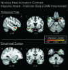
Migraine attack versus interictal state (n = 8 patients) contrast analysis of noxious heat (pain threshold +1°C) activation. Red-to-yellow voxels indicate areas with a significant contrast (determined by Gaussian mixture modeling) within the TP and the parahippocampal gyrus, as defined by the Harvard–Oxford Cortical Atlas as implemented by FSL. A minimum cluster criterion of 7 voxels in original space (0.30 cm3) was implemented to identify significant clusters. Blue circles highlight the TP and EC regions with significant contrasts in Experiment 1. Both the TP (maximum z-statistic: 2.23; volume: 0.57 cm3) and EC (maximum z-statistic: 2.57; volume: 0.54 cm3) show significantly increased activation during a migraine attack. A, anterior; C, contralateral; I, ipsilateral; and P, posterior.
Figure 5.
Figure 5.
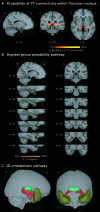
Group probability maps of white matter tracts connecting the TP and the pulvinar nucleus in the posterior thalamus based on the DTI tractography in 9 subjects. (A) Group probability of TP connectivity within pulvinar nucleus. The map represents the probability that voxels within the pulvinar nucleus have a connection to the TP (see Materials and Methods). (B) Group probability pathway connecting pulvinar nucleus and TP. The fact that there are common pulvinar voxels present in all the subjects with a maximum probability (yellow) strongly suggests that there is a connection between the TP and the pulvinar. The maps represent the probability of the presence of the pathways in at least 50% of the subjects to the maximum of presence in all subjects. (C) 3D rendering of the probabilistic pathway connecting TP and pulvinar nucleus. The probability map (red) is the same as shown in part (B). Pulvinar (green) and TP (olive) masks are shown here as well. Additionally, a projection from the pulvinar extends to the prefrontal cortex. A, anterior; C, contralateral; I, ipsilateral; and P, posterior.

References

    1. Afra J, Mascia A, Gerard P, Maertens de Noordhout A, Schoenen J. Interictal cortical excitability in migraine: a study using transcranial magnetic stimulation of motor and visual cortices. Ann Neurol. 1998;44:209–215. - PubMed
    1. Afra J, Proietti Cecchini A, Sandor PS, Schoenen J. Comparison of visual and auditory evoked cortical potentials in migraine patients between attacks. Clin Neurophysiol. 2000;111:1124–1129. - PubMed
    1. Afridi SK, Giffin NJ, Kaube H, Friston KJ, Ward NS, Frackowiak RS, Goadsby PJ. A positron emission tomographic study in spontaneous migraine. Arch Neurol. 2005;62:1270–1275. - PubMed
    1. Afridi SK, Matharu MS, Lee L, Kaube H, Friston KJ, Frackowiak RS, Goadsby PJ. A PET study exploring the laterality of brainstem activation in migraine using glyceryl trinitrate. Brain. 2005;128:932–939. - PubMed
    1. Ambrosini A, Rossi P, De Pasqua V, Pierelli F, Schoenen J. Lack of habituation causes high intensity dependence of auditory evoked cortical potentials in migraine. Brain. 2003;126:2009–2015. - PubMed

Publication types