. 2010 Jul 21;29(14):2290-300.
doi: 10.1038/emboj.2010.126. Epub 2010 Jun 18.

P2X4 receptors mediate PGE2 release by tissue-resident macrophages and initiate inflammatory pain

Affiliations

P2X4 receptors mediate PGE2 release by tissue-resident macrophages and initiate inflammatory pain

Lauriane Ulmann et al. EMBO J. .

Abstract

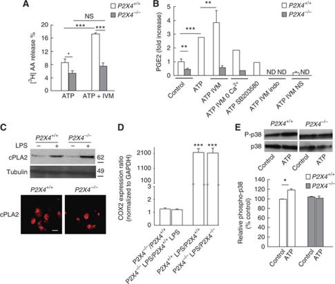
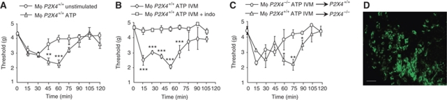
Prostaglandin E2 (PGE2) is a key mediator of inflammation and contributes to pain hypersensitivity by promoting sensory neurons hyperexcitability. PGE2 synthesis results from activation of a multi-step enzymatic cascade that includes cyclooxygenases (COXs), the main targets of non-steroidal anti-inflammatory drugs (NSAIDs). Although NSAIDs are widely prescribed to reduce inflammatory symptoms such as swelling and pain, associated harmful side effects restrict their long-term use. Therefore, finding new drugs that limit PG production represents an important therapeutic issue. In response to peripheral inflammatory challenges, mice lacking the ATP-gated P2X4 channel (P2X4R) do not develop pain hypersensitivity and show a complete absence of inflammatory PGE2 in tissue exudates. In resting conditions, tissue-resident macrophages constitutively express P2X4R. Stimulating P2X4R in macrophages triggers calcium influx and p38 MAPK phosphorylation, resulting in cytosolic PLA2 (cPLA2) activation and COX-dependent release of PGE2. In naive animals, pain hypersensitivity was elicited by transfer into the paw of ATP-primed macrophages from wild type, but not P2X4R-deficient mice. Thus, P2X4Rs are specifically involved in inflammatory-mediated PGE2 production and might therefore represent useful therapeutic targets.


Conflict of interest statement

The authors declare that they have no conflict of interest.

Figures

Figure 1
Figure 1
Pain behaviours in P2X4R-deficient mice. (A) Behavioural response to different amounts of 5% formalin injection in the hindpaw. Left panel, no significant difference in nocifensive behaviour was observed between the two genotypes when 20 μl of formalin was injected. Right panel, behavioural response to 15 μl injection was attenuated in WT mice, and P2X4R-deficient mice show a significant reduction of phase 2 behaviour. N=6 mice per group. (B) Mechanical hypersensitivity induced by injection of carrageenan (15 μl, 1% in saline) was measured 2 h after injection. WT mice develop a strong hyperalgesic response compared with baseline (P=0.0004), which is significantly reduced in P2X4−/− mice. N=6 animals per group. (C) Lack of long lasting inflammatory pain in P2X4R-deficient mice. Withdrawal threshold to mechanical stimulation was measured on injected side before and 24 h, 3 and 7 days after subcutaneous CFA injection in the paw. N=12 animals per group. (D) In the hot-plate test, there was no difference in latency between WT and P2X4−/− mice at 50°C. Note a slight hypersensitivity in P2X4−/− mice at higher temperatures, N=10 animals per group. (E) Direct injection of PGE2 in the paw induces mechanical hypersensitivity in P2X4−/− mice. Paw withdrawal threshold to mechanical stimulation was measured before, 30 and 60 min after PGE2 (100 ng, 10 μl) injection. N=6 mice per group. For all panels, results are expressed as mean±s.e.m., *P<0.05, **P<0.01, ***P<0.005, one-way ANOVA.
Figure 2
Figure 2
P2X4R-deficient mice show impaired peripheral PGE2 production in response to inflammatory challenge. PGE2 concentrations were measured in paw exudates in saline-injected animals, 2 h post-carrageenan injection (A) or 2 and 4 h after CFA injection (B). In both conditions, PGE2 concentrations were significantly reduced in paw exudates of P2X4R-deficient mice when compared with WT mice. Results were normalized to the mean concentration of PGE2 in WT saline group. N=6 mice per group, *P<0.05, ***P<0.005, Student t-test. (C) Mechanical allodynia was significantly reduced in P2X4R-deficient mice 2 and 4 h post-CFA injection. N=6 mice per group, *P<0.05, one-way ANOVA.
Figure 3
Figure 3
Paw-resident macrophages express P2X4R. (A) In paw sections of CX3CR1+/eGFP mice, P2X4R immunostaining colocalizes with eGFP (top row, scale bar=50 μm). Bottom row shows the colocalization of eGFP with the specific macrophage marker F4/80 at lower magnification (scale bar=100 μm). (B) Absence of recruitment of inflammatory cells by carrageenan in paw of CX3CR1+/eGFP mice. Macrophages were identified in control conditions and 2 h post-carrageenan injection by F4/80-positive staining (top row, scale bar=100 μm) and by the expression of eGFP (bottom row, scale bar=50 μm). (C) Western blot analysis shows that P2X4R expression in paw tissue is not different between control and treated mice. Each lane represents two animals. Experiment was repeated twice. (D, E) Carrageenan did not induce neutrophil recruitment. Neutrophils were identified in WT mice using GR1 immunostaining (D), or myeloperoxidase (MPO) assay (E), 2 h post-carrageenan or 24 h post-CFA. No recruitment was observed 2 h post-carrageenan, whereas it was clearly present 24 h after CFA injection. Scale bar=100 μm. ***P<0.005, Student t-test.
Figure 4
Figure 4
Peritoneal macrophages express functional P2X4R. (A) In WT peritoneal macrophages, P2X4R immunostaining (green) colocalized with that of F4/80 (red), whereas in P2X4R-deficient cell β-galactosidase (green) substitutes for the P2X4R immunostaining. Scale bar=10 μm. (B) Western blot analysis of P2X4R expression in peritoneal exudates or spleen tissue. Representative experiment out of three. (C, D) Characterization of P2X4R-induced calcium signals in peritoneal macrophages. (C) Representative recording of ATP-induced calcium signals in single WT and P2X4R-deficient peritoneal macrophages. ATP (20 μM) was applied for 10 s three times every 2 min and then a fourth application was made in the presence of ivermectin (IVM, 3 μM). The rundown of ATP-induced calcium signals was much more pronounced in P2X4R-deficient macrophages, and it was not reversed by IVM. (D) Population analysis of ATP-induced calcium signals as described in (C) and performed in the presence of A-740003 (10 μM) a specific P2X7 antagonist (hatched bars). Fluorescence ratios were normalized to the mean of F340/F380 ratio measured for the first ATP application. N>50 cells. *P<0.05, **P<0.01, ***P<0.005, one-way ANOVA.
Figure 5
Figure 5
P2X4R activation evokes arachidonic acid (AA) and PGE2 release from macrophages. (A) In WT macrophages, ATP (50 μM) induces [3H]AA release that is enhanced in the presence of IVM (3 μM), whereas in P2X4−/− macrophages ATP-evoked [3H]AA is reduced and IVM has no effect. N=5 independent experiments, one-way ANOVA, *P<0.05, ***P<0.005. (B) Stimulation of WT macrophages with ATP (50 μM) induced a three-fold increase of PGE2 release over baseline, which was further increased by IVM (3 μM). ATP-evoked PGE2 release was reduced in the absence of extracellular calcium and totally inhibited by pretreatment with p38 MAPK inhibitor SB203580 (10 μM), or with the COX inhibitors indomethacin (indo, 20 μM) or NS-398 (NS, 10 μM). Note that in P2X4−/− cells, basal PGE2 levels are reduced and that ATP/IVM do not trigger PGE2 release. To allow for inter-experiment comparisons, results were normalized to the mean concentration of PGE2 in WT control conditions, for each given experiment. N=3 independent experiments, one-way ANOVA, **P<0.01, ***P<0.005. ND, not detectable. (C) Western blot and immunocytochemistry analysis show that cPLA2 expression or its induction by LPS is not altered in P2X4−/− macrophages (scale bar=20 μm). N=2 experiments. (D) Transcriptional regulation of COX2 by LPS is not altered in P2X4−/− bone marrow-derived macrophages. Expression of COX2 mRNA was quantified in bone marrow-derived macrophages culture treated or not with LPS (1 μM, 6 h) from WT and P2X4R-deficient mice. Results are mean±s.e.m. of three independent experiments. One-way ANOVA, ***P<0.001. (E) Top panel, representative western blot analysis indicating that ATP (20 μM) induces an increase of P-p38 MAPK in WT but not in P2X4−/− macrophages. Bottom panel, quantification of N=3 independent experiments, one-way ANOVA, *P<0.05.
Figure 6
Figure 6
Transfer of ATP-primed macrophages induces P2X4-dependent pain hypersensitivity. (A) Peritoneal macrophages from CX3CR1+/eGFP mice were stimulated with ATP (50 μM) or left untreated and injected in the paw of WT mice; withdrawal thresholds to mechanical stimulation were measured every 15 min. Administration ATP-primed macrophages induced biphasic hypersensitivity, whereas that of unstimulated macrophages only induced a transient response. *P<0.05, **P<0.01, ***P<0.005, one-way ANOVA. (B) Similar experiments were performed with macrophages pretreated (or not) with indomethacin (20 μM) before wash and further stimulation with ATP (50 μM) and IVM (3 μM). Indomethacin completely abolished both phases of hypersensitivity. (C) ATP/IVM-primed macrophages from (CX3CR1+/eGFP X P2X4−/−) and CX3CR1+/eGFP mice were transferred to P2X4+/+ or P2X4−/− mice, respectively. Transfer of P2X4-deficient macrophages induced only transient hypersensitivity that returned to basal threshold values within 60 min. Transfer of CX3CR1+/eGFP macrophages in P2X4-deficient mice induced biphasic hypersensitivity, as observed in P2X4+/+ recipient mice N=4–8 animals per point. *P<0.05, **P<0.01, ***P<0.005, one-way ANOVA. All results are expressed as mean±s.e.m. (D) Transferred CX3CR1+/eGFP macrophages were still present in paw 6 h after injection. Scale bar=20 μm.

Similar articles

Cited by

References

    1. Avignone E, Ulmann L, Levavasseur F, Rassendren F, Audinat E (2008) Status epilepticus induces a particular microglial activation state characterized by enhanced purinergic signaling. J Neurosci 28: 9133–9144 - PMC - PubMed
    1. Balboa MA, Balsinde J, Johnson CA, Dennis EA (1999) Regulation of arachidonic acid mobilization in lipopolysaccharide-activated P388D(1) macrophages by adenosine triphosphate. J Biol Chem 274: 36764–36768 - PubMed
    1. Bhattacharyya S, Gill R, Chen ML, Zhang F, Linhardt RJ, Dudeja PK, Tobacman JK (2008) Toll-like receptor 4 mediates induction of the Bcl10-NFkappaB-interleukin-8 inflammatory pathway by carrageenan in human intestinal epithelial cells. J Biol Chem 283: 10550–10558 - PMC - PubMed
    1. Block ML, Hong JS (2005) Microglia and inflammation-mediated neurodegeneration: multiple triggers with a common mechanism. Prog Neurobiol 76: 77–98 - PubMed
    1. Bonventre JV, Sapirstein A (2002) Group IV cytosolic phospholipase A2 (PLA2) function: insights from the knockout mouse. Adv Exp Med Biol 507: 25–31 - PubMed

Publication types

MeSH terms