. 2010 Jul 21;29(14):2315-28.
doi: 10.1038/emboj.2010.133. Epub 2010 Jun 18.

Cooperative interactions at the SLP-76 complex are critical for actin polymerization

Affiliations

Cooperative interactions at the SLP-76 complex are critical for actin polymerization

Mira Barda-Saad et al. EMBO J. .

Abstract

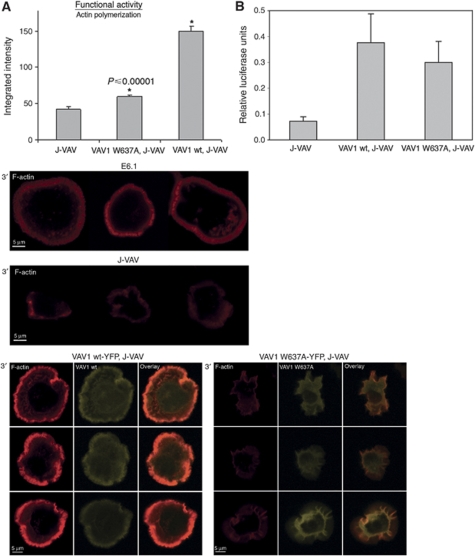
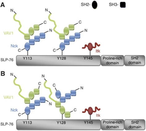
T-cell antigen receptor (TCR) engagement induces formation of multi-protein signalling complexes essential for regulating T-cell functions. Generation of a complex of SLP-76, Nck and VAV1 is crucial for regulation of the actin machinery. We define the composition, stoichiometry and specificity of interactions in the SLP-76, Nck and VAV1 complex. Our data reveal that this complex can contain one SLP-76 molecule, two Nck and two VAV1 molecules. A direct interaction between Nck and VAV1 is mediated by binding between the C-terminal SH3 domain of Nck and the VAV1 N-terminal SH3 domain. Disruption of the VAV1:Nck interaction deleteriously affected actin polymerization. These novel findings shed new light on the mechanism of actin polymerization after T-cell activation.


Conflict of interest statement

The authors declare that they have no conflict of interest.

Figures

Figure 1
Figure 1
SLP-76, Nck and VAV1 complex formation. Sedimentation coefficient distributions ck(s) of the SLP-76 complexes formed in the presence of Nck and/or VAV1. The data are from sedimentation velocity experiments with different mixtures, of SLP-76 peptides pY113pY145 (squares), pY128pY145 (triangles) or pY113pY128pY145 (circles), all at 1.5 μM, in the presence of 6 μM VAV1 (blue), 6 μM Nck (green) or both (magenta). All data shown with symbols and/or solid lines are cSLP(s) distributions of SLP-76 peptides and its complexes only, calculated by exploiting a spectrally distinct chromophoric label. In the presence of protein, the peptides track the sedimentation of complexes at s-values >2 S. (A) The control sedimentation coefficient distribution of SLP-76 peptides alone is shown as black solid line. The result of a control experiment with VAV1 alone is shown as blue dotted line, which displays cVAV(s) calculated by taking advantage of its own characteristic spectral properties of the FAM label on VAV1. In the presence of the triply phosphorylated peptide, a population of 4.2 S complexes (likely in 2:1 stoichiometry) can be discerned. (B) VAV1 and Nck can both simultaneously bind to SLP-76 pY113,145, as can be discerned from the shift towards faster-sedimenting complexes in the presence of VAV1+Nck. The sedimentation coefficient distribution from an experiment with Nck alone, as observed through its own specific spectral signature cNck(s), is shown as dotted line. (C) Higher-order complexes are formed on the triply phosphorylated peptide in the presence of VAV1+Nck. For the experiment with both proteins (magenta), the dotted lines show the sedimentation coefficient distributions calculated from multi-signal analysis for VAV1-containing complexes cVAV(s) (blue dotted line) and Nck-containing complexes cNck(s) (green dotted line) in the same solution.
Figure 2
Figure 2
FRET analysis of the triple molecular complex SLP-76, Nck and VAV1. (A) SLP-76-deficient Jurkat J14 cells were reconstituted with YFP-SLP-76 and transfected with CFP-Nck. VAV1-deficient Jurkat J-VAV cells were cotransfected with CFP-SLP-76 and YFP-Nck. High FRET efficiency was observed in the presence, as well as in the absence, of VAV1 (P⩽0.094). (B) J14 cells expressing YFP-SLP-76 CFP-Nck were transiently transfected with double-stranded siRNA oligonucleotides comprising Nckα&β SH2 domain-specific sequences or a non-specific sequence (negative control). At 48 h after transfection, cells were analysed as described above for protein levels of Nck using anti-Nck antibody. The siRNA-mediated Nck downregulation was compared with the level of GAPDH. (C) J14 cells were reconstituted with YFP-SLP-76 and transfected with VAV1-CFP. The decrease in FRET efficiency using Nckα&β siRNA shows that Nck is essential for the recruitment of VAV1 to SLP-76 signalling complexes (P⩽0.000015). The results are based on more than three independent experiments.
Figure 3
Figure 3
The interaction between Nck and VAV1 is independent of SLP-76. (A) FRET analysis of the molecular interaction between Nck and VAV1 in Jurkat E6.1 T cells expressing YFP-Nck VAV1-CFP was performed in comparison to SLP-76-deficient Jurkat J14 cells (P⩽0.07) or J14 reconstituted with SLP-76 wt (P⩽0.57) expressing VAV1-YFP CFP-Nck. No significant difference in the FRET efficiency was measured between Nck and VAV1 expressed in J14 to J14 reconstituted with wtSLP-76 cells (P⩽0.412). Different cell panels are presented. The results are based on more than five independent experiments. (B) Immunoprecipitation assay of the interaction between Nck and VAV1 in the presence of SLP-76, E6.1 Jurkat cells, and in the absence of SLP-76, J14 Jurkat cells. Three independent experiments were performed. One representative experiment is shown. (C) J14 cells were reconstituted with SLP-76 wt or SLP-76Y3F mutant form. Immunoprecipitation was performed with anti-VAV1, and the membrane was immunoblotted with anti-VAV1 and anti-Nck. (D) Nck and VAV1 interaction was confirmed in PBLs. The molecular interaction was detected in unstimulated as well as TCR-stimulated cells. Six independent experiments were performed. One representative experiment is shown. (E) Samples from stimulated and unstimulated PBLs were analysed by western blotting for pSLP-76 levels using anti-phosphotyrosine antibody.
Figure 4
Figure 4
Direct interaction between VAV1 and Nck is mediated by the C-terminal SH3 domain of Nck. (A) FRET analysis of the association of Nck wt with VAV1 was determined in J14 cells expressing VAV1-CFP YFP-Nck versus J14 cells expressing Nck mutated at the C-terminal SH3 domain. Different cell panels are presented. (B) Mutations at the N- or middle-SH3 domain of Nck were examined as well and compared with the Nck wt. The cells were plated on stimulatory coverslip and fixed after 1, 3 or 5 min. Three independent experiments were performed. (C) Immunoprecipitation assay of the interaction between VAV1 wt and Nck wt in comparison to VAV1 wt and mutant form of Nck mutated at C-terminal SH3 domain, Nck*SH3(C) expressed in J14. More than three independent experiments were performed. One representative experiment is shown.
Figure 5
Figure 5
The N-terminal SH3 domain of VAV1 is required for a direct association with Nck. (A) FRET analysis of the association of VAV1 with Nck in J14 cells was performed and compared between VAV1 wt-YFP and VAV1 W637A-YFP. The results are based on three independent experiments. (B) Analysis of VAV1-deficient cells, J-VAV, stably expressing VAV1 wt-YFP or VAV1 W637A-YFP for their VAV1 protein expression was performed. Cells were lysed. The whole-cell lysates were resolved on SDS–PAGE and probed with anti-VAV1 antibodies to determine protein level expression. (C) Immunoprecipitation experiments were performed to detect the interaction between Nck to VAV1 wt or mutated VAV1 W637A form in J-VAV cells. Six independent experiments were performed. One representative experiment is shown. (D) The interaction between SLP-76 to VAV1 was detected by immunoprecipitation using VAV1 wt-YFP versus VAV W637A-YFP expressed in J-VAV cells. Immunoprecipitation of SLP-76 and immunoblotting with anti-VAV1 and anti-Nck antibodies were performed.
Figure 6
Figure 6
The N-terminal SH3 domain of VAV1 is necessary for an efficient actin polymerization. (A) J-VAV cells were reconstituted with VAV1 W637A mutant or VAV1 wt. Cells were seeded over a stimulatory coverslip pre-coated with anti-CD3 antibody then fixed 5 min into the spreading process and their actin polymerization measured by phalloidin staining. The integration of fluorescence intensity of cells labelled with phalloidin is presented. The results were compared with J-VAV cells. Different cell panels are presented. Three independent experiments were performed. (B) J-VAV cell lines stably expressing VAV1 wt or VAV1 W637A mutant or vector were transiently cotransfected with an NFAT luciferase reporter plasmid. After 16 h, the cells were incubated for 6 h in tissue culture medium alone (unstimulated), with plate-bound monoclonal antibody to CD3, or with a combination of phorbol 12-myristate 13-acetate (PMA) and ionomycin. Cells were then lysed and assayed for luciferase activity as described in Materials and methods. All values were normalized to internal Renilla controls. The results are expressed as a fraction of the activity obtained on stimulation with PMA and ionomycin. No significant difference was found between VAV1 wt and VAV1 W637A mutant cells (P⩽0.59). The results are the average of five independent experiments.
Figure 7
Figure 7
Schematic representation of possible oligomerization models of SLP-76-binding sites (A, B). On TCR activation, SLP-76 is fully phosphorylated. This event leads to the oligomerization of SLP-76 by Nck and VAV1 and possibly Itk-containing complexes. The oligomerized SLP-76 molecules then serve as a nucleation site for other signalling molecules, resulting in the activation of intracellular signalling pathways.

Similar articles

Cited by

References

    1. Astoul E, Laurence AD, Totty N, Beer S, Alexander DR, Cantrell DA (2003) Approaches to define antigen receptor-induced serine kinase signal transduction pathways. J Biol Chem 278: 9267–9275 - PubMed
    1. Badour K, Zhang J, Shi F, McGavin MK, Rampersad V, Hardy LA, Field D, Siminovitch KA (2003) The Wiskott-Aldrich syndrome protein acts downstream of CD2 and the CD2AP and PSTPIP1 adaptors to promote formation of the immunological synapse. Immunity 18: 141–154 - PubMed
    1. Balbo A, Minor KH, Velikovsky CA, Mariuzza RA, Peterson CB, Schuck P (2005) Studying multiprotein complexes by multisignal sedimentation velocity analytical ultracentrifugation. Proc Natl Acad Sci USA 102: 81–86 - PMC - PubMed
    1. Barda-Saad M, Braiman A, Titerence R, Bunnell SC, Barr VA, Samelson LE (2005) Dynamic molecular interactions linking the T cell antigen receptor to the actin cytoskeleton. Nat Immunol 6: 80–89 - PubMed
    1. Billadeau DD, Burkhardt JK (2006) Regulation of cytoskeletal dynamics at the immune synapse: new stars join the actin troupe. Traffic 7: 1451–1460 - PMC - PubMed

Publication types

MeSH terms