. 2010 Jul;17(7):822-9.
doi: 10.1038/nsmb.1854. Epub 2010 Jun 20.

Ion/substrate-dependent conformational dynamics of a bacterial homolog of neurotransmitter:sodium symporters

Affiliations

Ion/substrate-dependent conformational dynamics of a bacterial homolog of neurotransmitter:sodium symporters

Derek P Claxton et al. Nat Struct Mol Biol. 2010 Jul.

Abstract

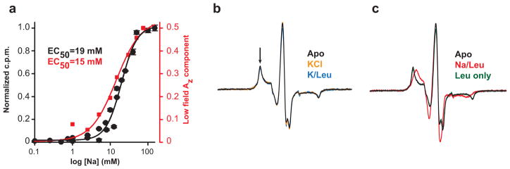
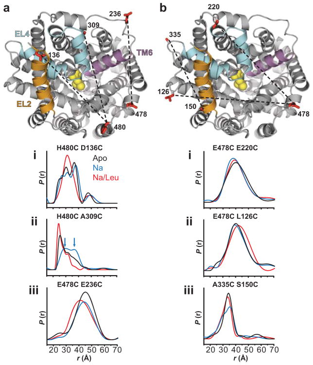
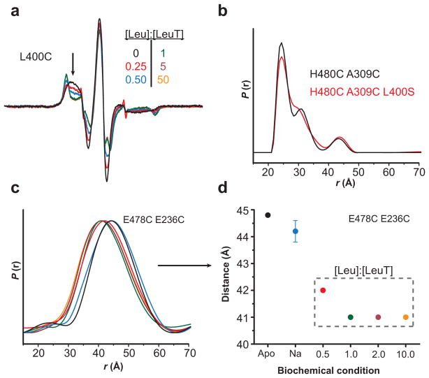
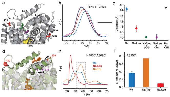
Crystallographic, computational and functional analyses of LeuT have revealed details of the molecular architecture of Na(+)-coupled transporters and the mechanistic nature of ion/substrate coupling, but the conformational changes that support a functional transport cycle have yet to be described fully. We have used site-directed spin labeling and electron paramagnetic resonance (EPR) analysis to capture the dynamics of LeuT in the region of the extracellular vestibule associated with the binding of Na(+) and leucine. The results outline the Na(+)-dependent formation of a dynamic outward-facing intermediate that exposes the primary substrate binding site and the conformational changes that occlude this binding site upon subsequent binding of the leucine substrate. Furthermore, the binding of the transport inhibitors tryptophan, clomipramine and octyl-glucoside is shown to induce structural changes that distinguish the resulting inhibited conformation from the Na(+)/leucine-bound state.


Conflict of interest statement

Author Information

The authors have no competing financial interests.

Figures

Figure 1
Figure 1
Na+ binding establishes an outward-facing conformation and Leu binding closes access to the extracellular vestibule. Representative EPR spectra (a) and the corresponding NiEDDA accessibility profiles (b) in proteoliposomes demonstrate that increased water penetration correlates with increased spin label dynamics (arrows, panel a). Leu binding leads to a decrease in spin label dynamics which corresponds with a decrease in solvent accessibility. The low field changes in EPR lineshape for V33C are highlighted in the boxed inset in a. Data in (b) are shown as mean ± S.D from three independent measurements. (c,d) Residue-specific mapping of the change in NiEDDA accessibility upon Na+ binding (relative to the Apo state) and Leu binding (relative to the Na+ state) onto the LeuT crystal structure (PDB 2A65). Na+ ions are depicted as green spheres and Leu is colored yellow in a space-filling model. Positions exhibiting an increase in accessibility are shaded in blue according to the color scale. Structures were generated using UCSF Chimera.
Figure 2
Figure 2
Changes in the EPR spectrum are dependent upon Na+ binding. (a) The [Na+]-dependent increase in probe mobility at 111C (Figure 1a) as monitored by the intensity of the low field Az component (arrow in b) revealed an EC50Na+ similar to that obtained by Na+-stimulated binding of 100 nM 3H-Leu. (b–c) Leu-dependent changes in the EPR spectrum at I111C required the presence of Na+. In the absence of Na+ or the presence of K+, Leu did not induce changes in the EPR spectrum relative to the Apo state.
Figure 3
Figure 3
Substrate binding induces dehydration of the S1 site. (a) Representation of the LeuT crystal structure (PDB 2A65) highlighting the bound Leu in the S1 site (Leu is shown in a yellow space-filling model), the position of a key residue, F253 (red stick representation), and the distribution of water molecules (shown as blue spheres). Note that there are no water molecules in the S1 site. (b) Na+-dependent Ala uptake (Methods) was greatly impaired in the F253C mutant. (c) Na+-dependent binding of 100 nM 3H-Leu was reduced ~50% for the F253C mutant relative to WT. Data shown are mean ± S.E.M. (d) Accessibility to both 50mM NiEDDA and 20% O2 of F253C in proteoliposomes was low in all states, consistent with a buried location. Equimolar Leu binding decreased the EPR spectral breadth at both room temperature (e) and –53°C (f) indicating that water becomes excluded from the primary binding site (S1). The structure in (a) was generated using PyMOL.
Figure 4
Figure 4
Distance measurements reveal Na+- and Leu-dependent spatial rearrangements. (a) Na+ binding enhanced an outward-facing conformation characterized by movement of EL4 as suggested by the H480C/A309C pair distance measures. Blue arrows in the distance distribution of H480C/A309C indicate two unresolved distance populations in the Na+ intermediate. Leu binding to the Na+ intermediate decreased the distance between the probes for both H480C/A309C and E478C/E236C pairs. Furthermore, Leu binding increased structural homogeneity as indicated by a narrowing of the distance distribution for both H480C/D136C and H480C/A309C pairs. (b) Limited distance changes were observed for pairs sampling protein structure outside of the vestibule. The LeuT structure was obtained from PDB 2A65. The structures were generated using PyMOL.
Figure 5
Figure 5
Leu binding to the S1 site is the primary determinant of Leu-dependent conformational changes. (a) The decrease in spin label mobility at L400C was dependent on the [Leu]. The Leu titration of LeuT-L400C indicated a maximum change in EPR lineshape (arrow) at a 1:1 molar ratio of Leu-to-LeuT. Because the L400C mutation disrupts binding to the S2 site, these spectral changes result from binding in the S1 site. (b) The distance distributions of LeuT-H480C/A309C (2:1 Leu-to-LeuT stoichiometry) and –H480C/A309C/L400S (1:1 stoichiometry) are superimposable, suggesting that the Leu-dependent change in distance is due to Leu occupancy in the S1 site. (c–d) The change in distance between LeuT-E478C/E236C (2:1 stoichiometry) is saturated at a 1:1 ratio, consistent with S1-driven conformational changes. The distances in (d) were obtained from the peak probability of the distance distributions in (c).
Figure 6
Figure 6
Binding of inhibitors lead to structural changes distinct from the Na+/Leu bound conformation. (a) Both OG (green stick) and CMI (purple stick) can occupy the extracellular vestibule with Leu (yellow sphere) in the S1 site. For clarity, OG (from PDB 3GJC) and CMI (from PDB 2Q6H) have been mapped onto the Na+/Leu bound LeuT crystal structure (PDB 2A65). (b–c) OG and CMI binding produce similar decreases in distance between the probes. Addition of 1mM CMI to LeuT in the presence of Na+ alone did not shift the distance distribution. The mean ± S.D. of three repeated distances measurements in (c) were obtained from the peak probability of the distance distributions in (b). (d) Trp (orange stick) competitively inhibits transport by binding to the S1 site and displacing Leu. (e) 20mM Trp increases the average distance between H480C/A309C, selectively enhancing the outer-most component of the Na+ intermediate (gray dashed box). For comparison, the Trp-bound LeuT crystal structure (green, PDB 3F3A) overlays the Na+/Leu crystal structure (gray, PDB 2A65) illustrating the altered conformation EL4 adopts in the presence of Trp. Arrows in (d) indicate the position change of A309 and A315 in the LeuT-Trp structure. (f) Trp binding doubles the NiEDDA accessibility of the A315C mutant in proteoliposomes (relative to the Na+-bound intermediate), consistent with altering the conformation of EL4. Data are shown as mean ± S.D. The structures in (a) and (d) were generated using PyMOL
Figure 7
Figure 7
Configuration changes of the TM3–TM6 aromatic cluster associated with the conformational transition simulated in the 660 ns MD trajectory. The starting conformation was the LeuT crystal structure, in which the Leu molecule found in the S1 site (PDB 2A65) was removed (a) and (b) are the crystal structures with substrate (Leu) or inhibitor (Trp) bound in the S1 site, respectively. Y107 and Y108 from TM3, and F252 and F253 from TM6 are rendered in sticks. The ligands are in space-filling representation. (c) The outward movement of TM6 (with Na+ only bound) indicated by a white arrow, overlaps the one evident in the LeuT-Trp structure (PDB 3F3A), which has been proposed to represent the ‘open-to-out’ conformation. (d) Superimposed view of the aromatic cluster in the context of the Na1 site. The side chains are colored corresponding to the backbone colors in ac. The overlap between F253 (in the Na-only simulation) and the Trp in the extracellular vestibule of the Trp-bound crystal structure (Trp602 of PDB 3F3A), is indicated by a dotted circle. Only the ligand and Na+ ions from the Trp-bound structure are shown. (e) Changes in Χ1 rotamer values for the aromatic cluster residues along the MD trajectory.

References

    1. Nelson N. The family of Na+/Cl− neurotransmitter transporters. J Neurochem. 1998;71:1785–803. - PubMed
    1. Wadiche JI, Kavanaugh MP. Macroscopic and microscopic properties of a cloned glutamate transporter/chloride channel. J Neurosci. 1998;18:7650–61. - PMC - PubMed
    1. Amara SG, Sonders MS. Neurotransmitter transporters as molecular targets for addictive drugs. Drug Alcohol Depend. 1998;51:87–96. - PubMed
    1. Rudnick G, editor. Mechanisms of biogenic amine neurotransmitter transporters. Humana Press Inc; Totowa, New Jersey: 2002. pp. 25–52.
    1. Krause S, Schwarz W. Identification and selective inhibition of the channel mode of the neuronal GABA transporter 1. Mol Pharmacol. 2005;68:1728–35. - PubMed

Publication types

MeSH terms