. 2010 Aug 26;29(34):4848-58.
doi: 10.1038/onc.2010.235. Epub 2010 Jun 21.

GLI3-dependent repression of DR4 mediates hedgehog antagonism of TRAIL-induced apoptosis

Affiliations

GLI3-dependent repression of DR4 mediates hedgehog antagonism of TRAIL-induced apoptosis

S Kurita et al. Oncogene. .

Abstract

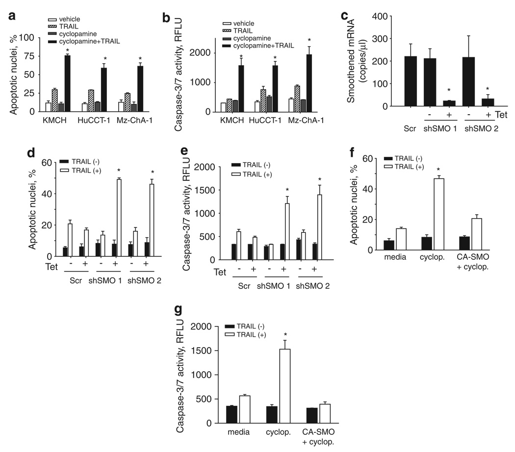
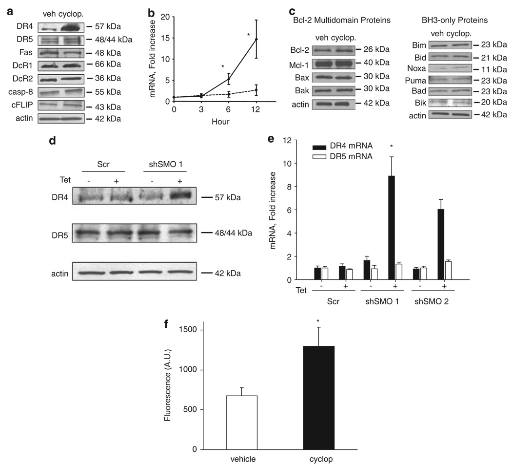
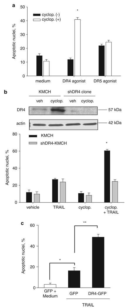
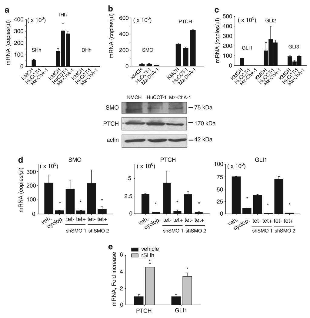
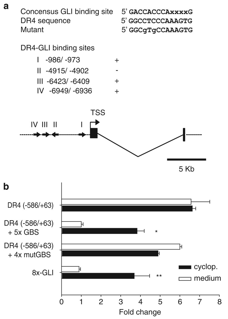
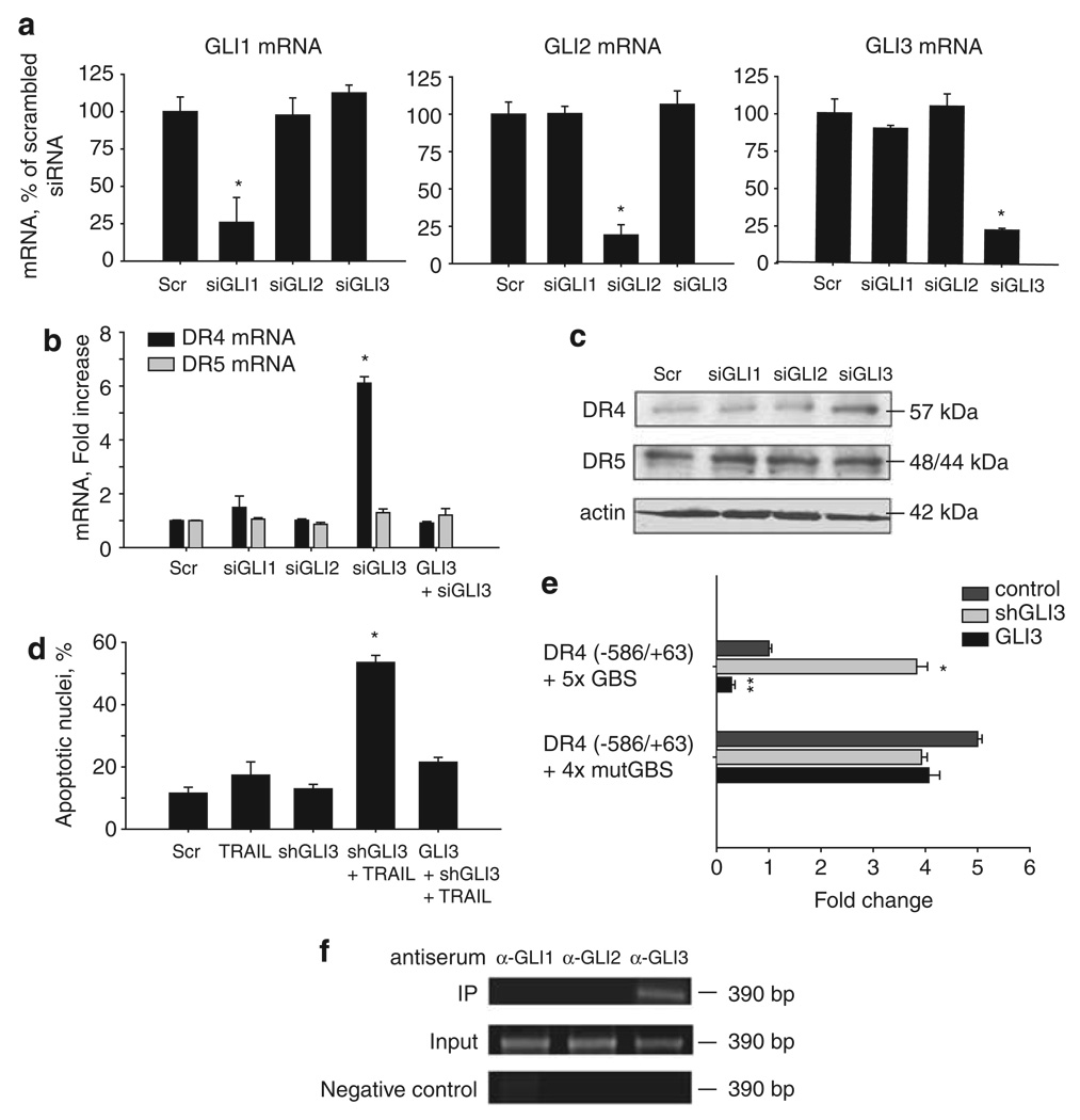
Tumor necrosis factor-related apoptosis-inducing ligand (TRAIL) induces apoptosis through its cognate receptors death receptor 4 (DR4) and death receptor 5 (DR5), preferentially in malignant cells. However, many malignant cells remain resistant to TRAIL cytotoxicity by poorly characterized mechanisms. Here, using cholangiocarcinoma cells, as a model for TRAIL resistance, we identified a role for the oncogenic Hedgehog (Hh)-GLI pathway in the regulation of TRAIL cytotoxicity. Blockade of Hh using pharmacological and genetic tools sensitizes the cells to TRAIL cytotoxicity. Restoration of apoptosis sensitivity coincided with upregulation of DR4 expression, while expression of other death effector proteins remained unaltered. Knockdown of DR4 mimics Hh-mediated resistance to TRAIL cytotoxicity. Hh regulates the expression of DR4 by modulating the activity of its promoter. Luciferase, chromatin immunoprecipitation and expression assays show that the transcription factor GLI3 binds to the DR4 promoter and Hh requires an intact GLI3-repression activity to silence DR4 expression. Finally, small interfering RNA (siRNA)-targeted knockdown of GLI3, but not GLI1 or GLI2, restores DR4 expression and TRAIL sensitivity, indicating that the Hh effect is exclusively mediated by this transcription factor. In conclusion, these data provide evidence of a regulatory mechanism, which modulates TRAIL signaling in cancer cells and suggest new therapeutic approaches for TRAIL-resistant neoplasms.


Conflict of interest statement

Conflict of interest

The authors declare no conflict of interest.

Figures

Figure 1
Figure 1
Cyclopamine sensitizes cholangiocarcinoma cells to Tumor necrosis factor-related apoptosis-inducing ligand (TRAIL)-mediated cytotoxicity. (a) Cells were pretreated with vehicle (ethanol, 0.01% v/v) or cyclopamine (5 µm) for 24 h. After pretreatment, human recombinant TRAIL was added where indicated at 5 ng/ml in fresh medium and the cells were incubated for an additional 5 h. Cells were then stained with 4′,6-diamino-2-phenylindole dihydrochloride (DAPI) and cells with apoptotic morphology were counted. Mean ± s.e.m., *P<0.001. (b) In parallel, cells were pretreated with vehicle or cyclopamine and treated with TRAIL as in panel (a) but after 6 h caspase-3/7 activity was measured. Mean ± s.e.m., *P<0.001. (c) KMCH cells were transfected with either of two independent short hairpin RNA (shRNA) expression vectors targeting Smoothened (shSMO 1 and shSMO 2) or a scrambled shRNA vector, followed by quantitative RT–PCR for Smoothened using total RNA. Results expressed as copies of each complementary DNA. Mean ± s.e.m., *P<0.001. (d) Cells transfected as in panel (c) were then treated with TRAIL (5 ng/ml) for 6 h, then stained with DAPI and cells with apoptotic morphology were counted. (e) Caspase-3/7 activity was measured 6 h after TRAIL treatment of cells transfected as in panel (c). Mean ± s.e.m., *P<0.001. (f, g) KMCH cells were pretreated with vehicle or cyclopamine as above, or transfected with a constitutively active Smoothened vector (CA-SMO). Cells were then stained with DAPI and cells with apoptotic morphology were counted (f) or caspase-3/7 activity was measured 6 h after TRAIL treatment (g). Mean ± s.e.m., *P<0.001.
Figure 2
Figure 2
Cyclopamine upregulates death receptor 4 (DR4) expression. (a) Immunoblotting of cell lysates from KMCH cells treated with vehicle or cyclopamine (5 µm) for Fas, TRAIL receptors DR4, DR5, DcR1 and DcR2, and proximal signaling proteins caspase 8 and cellular FLICE-inhibitory protein. Actin was used as a loading control. (b) Quantitative RT–PCR for DR4 and DR5 using total RNA from KMCH cells after treatment with cyclopamine for the indicated times. Expression was normalized to 18S rRNA levels and presented as fold increase over time. Mean ± s.e.m., *P<0.01. (c) Whole-cell lysates of KMCH cells treated with vehicle or cyclopamine (5 µm) were prepared for immunoblotting for B-cell leukemia 2family proteins, including BH3-only proteins. (d) KMCH cells transfected with scrambled control short hairpin RNA (shRNA) or two shRNA sequences targeting Smoothened (SMO shRNA 1 and SMO shRNA 2) were treated with tetracycline (1 mg/ml) to induce shRNA expression and whole-cell lysates prepared for immunoblotting. (e) In parallel, total RNA was prepared from KMCH cells transfected as indicated and RT–PCR for DR4 or DR5 was performed. Mean ± s.e.m., presented as fold increase over scrambled. *P<0.01. (f) DR4 surface expression was performed by using confocal microscopy. KMCH cells treated with cyclopamine (5 µm) for 24 h. Quantification of DR4-green fluorescent protein (GFP) localization was accomplished using the LSM210 imaging software. Fluorescence intensity for the green channel was quantified. Mean ± s.e.m. *P<0.05.
Figure 3
Figure 3
Death receptor 4 (DR4) receptor activation is necessary and sufficient for apoptosis in cyclopamine-sensitized cells. (a) Cells were pretreated with vehicle or cyclopamine (5 µm) for 24 h. DR4 agonist (0.1 µg/ml) or DR5 agonist (1 µg/ml) was then added in fresh medium and the cells were further incubated for the indicated time-intervals. Cells were then stained with 4′,6-diamino-2-phenylindole dihydrochloride (DAPI) and cells with apoptotic morphology were counted. Mean ± s.e.m., *P<0.001. (b) KMCH cells stably transfectred with DR4-shRNA and parent KMCH cells were incubated with vehicle or 5 µm cyclopamine for 24 h. Upper, Immunoblotting of whole cell lysates was performed using antibody against DR4. Actin was used as a loading control. Lower, Cells were pretreated where indicated with cyclopamine then treated with TRAIL (5 ng/ml) for 5 h followed by DAPI staining. Cells with apoptotic morphology were counted. Mean ± s.e.m., *P<0.001 compared with parent cells treated with TRAIL and cyclopamine. (c) KMCH cells transiently transfected with green fluroscent protein (GFP) (control) or DR4-GFP were treated where indicated with TRAIL (5 ng/ml) for 6 h. The number of GFP-positive cells with apoptotic nuclear morphology is presented as a percent of total GFP-positive cells. Data are mean ± standard error, *P<0.05; **P<0.01.
Figure 4
Figure 4
Cholangiocarcinoma cell lines expressed Hedgehog pathway mediators and respond to Hedgehog pathway activation and inhibition. (a) Quantitative RT–PCR for Sonic Hedgehog (Shh), Indian Hedgehog (Ihh), and Desert Hedgehog (Dhh) using total RNA from KMCH, HuCCT-1 and Mz-ChA-1 cells. Results are expressed as copies of each complementary DNA (cDNA) (Mean ± s.e.m.). (b) Upper, Quantitative RT–PCR for Smoothened and Patched-1 using total RNA from KMCH, HuCCT-1 and Mz-ChA-1 cells. Results are expressed as copies of each cDNA (Mean ± s.e.m.). Lower, Whole cell lysates from KMCH, HuCCT-1 and Mz-ChA-1 cells were probed using a Smoothened or a Patched-1 antibody. Actin was used as a loading control. (c) Quantitative RT–PCR for Hedgehog transcription factors, GLI1, -2 and -3, using total RNA from KMCH, HuCCT-1 and Mz-ChA-1 cells. Results expressed as copies of each cDNA (Mean ± s.e.m.). (d) Quantitative RT–PCR for Smoothened, Patched-1 and GLI1 using total RNA from KMCH cells after treatment with cyclopamine or gene silencing of Smoothened by a tetracycline-inducible shRNA technique. Results are expressed as copies of each cDNA (Mean ± s.e.m.). Cells were treated with cyclopamine 5 µm for 24 h before isolating mRNA or were transfected with Smoothened shRNA for 5 days before isolating mRNA. *P<0.001. (e) Quantitative RT–PCR for Patched-1, and GLI1 using total RNA from KMCH cells after treatment with recombinant sonic hedgehog ligand (rSHh). Results were normalized to 18S and expressed as fold increase over vehicle. Mean ± s.e.m., *P<0.01.
Figure 5
Figure 5
GLI3 directly repressed death receptor 4 (DR4) promoter function. (a) Putative GLI-binding sites in the DR4 promoter region. Nucleotide positions (I, II, III and IV) were counted from the transcription start site (TSS). Potential binding sites were observed in both the forward and the reverse direction; (+, sense; −, anti-sense) as indicated also by arrow on the schematic. (b) A DR4 promoter fragment (−586/+63) in pGL3, the same promoter plus five copies of the GLI-binding sequence (DR4 (−586/+63) + 5 × GBS), and a reporter construct containing 8 × GLI-binding sites (8 × -GLI) were used to evaluate the promoter activity on cyclopamine treatment. KMCH cells were co-transfected with 25 ng of pRL-CMV and 0.5 µg of indicated pGL3-based DR4 promoter reporter plasmids or 8 × -GLI reporter plasmids. Twenty-four hours after transfection, medium or cyclopamine (5 µm) was added where indicated for 24 h. Both firefly and Renilla luciferase activities were quantified and data (firefly/Renilla luciferase activity) are expressed as fold increase over DR4 promoter-reporter constructs containing 5 × GLI-binding sites treated with medium. Mean ± s.e.m., *P<0.001, **P<0.05.
Figure 6
Figure 6
Gene silencing of GLI3 sensitizes cholangiocarcinoma cells to TRAIL-mediated apoptosis through death receptor 4 (DR4) upregulation. (a) Quantitative RT–PCR for GLI1, -2 and -3 using total RNA from KMCH cells transfected with the specific siRNA for GLI1, -2 or -3. Forty-eight hours after transfection, total RNA was isolated from cells. Cells transfected with scramble small interfering RNA (siRNA) were used as a control. Results normalized to 18S and expressed as percentage of scrambled. Mean ± s.e.m., *P<0.001. (b) Quantitative RT–PCR for DR4 and DR5 using total RNA from KMCH cells after the specific GLI-siRNA transfection. Forty-eight hours after transfection, total RNA was isolated from cells. Cells transfected with scramble siRNA were used as a control. Results normalized to 18S and expressed as fold increase over scramble. GLI3 silencing was rescued where indicated by co-transfection with a GLI3 expression plasmid resistant to the siRNA. Mean ± s.e.m., *P<0.001. (c) Immunoblotting of whole cell lysates obtained from KMCH cells transfected with the specific siRNA targeting GLI1, -2, or -3. Seventy-two hours after transfection, total cellular protein was isolated from the cells. Membranes were probed using a DR4 and a DR5 antibody. Actin was used as a loading control. (d) KMCH cells transfectred with GLI3 shRNA or co-transfected with shGLI3 plus a GLI3-expression-plasmid resistant to the shRNA were incubated with 5 ng/ml human recombinant TRAIL for 5 h. Cells were then stained with 4′,6-diamino-2-phenylindole dihydrochloride (DAPI) and cells with apoptotic morphology were counted. Mean ± s.e.m., *P<0.001 compared with vehicle. (e) DR4 promoter constructs (−586/+63) with a tandem repeat of the candidate GLI-binding site (DR4 (−586/+63) + 5 × GBS) or a tandem repeat of a mutated GLI-binding site (DR4 (−586/+63) + 4 × mutGBS) were assayed in KMCH cells. Cells were co-transfected with shGLI3 or a GLI3-expression construct as indicated. Mean ± s.e.m. *P<0.001 compared with medium; **P<0.005 compared with medium. (f) Chromatin immunoprecipitation was performed on KMCH cells using anti-GLI1, -GLI2 or –GLI3 anti-serum and the pull down used for DR4 promoter polymerase chain reaction using primers flanking the candidate GLI-binding site I. The polymerase chain reaction amplicon was visualized by ethidium bromide staining after agarose electrophoresis.

References

    1. Akazawa Y, Mott JL, Bronk SF, Werneburg NW, Kahraman A, Guicciardi ME, et al. Death receptor 5 internalization is required for lysosomal permeabilization by TRAIL in malignant liver cell lines. Gastroenterology. 2009;136:2365–2376. e2361–e2367. - PMC - PubMed
    1. Ashkenazi A, Dixit VM. Apoptosis control by death and decoy receptors. Curr Opin Cell Biol. 1999;11:255–260. - PubMed
    1. Berman DM, Karhadkar SS, Maitra A, Montes De Oca R, Gerstenblith MR, Briggs K, et al. Widespread requirement for Hedgehog ligand stimulation in growth of digestive tract tumours. Nature. 2003;425:846–851. - PubMed
    1. Bigelow RL, Chari NS, Unden AB, Spurgers KB, Lee S, Roop DR, et al. Transcriptional regulation of bcl-2 mediated by the sonic hedgehog signaling pathway through gli-1. J Biol Chem. 2004;279:1197–1205. - PubMed
    1. Chen JK, Taipale J, Cooper MK, Beachy PA. Inhibition of Hedgehog signaling by direct binding of cyclopamine to Smoothened. Genes Dev. 2002;16:2743–2748. - PMC - PubMed

Publication types

MeSH terms

Substances