. 2010 Sep 2;29(35):4925-37.
doi: 10.1038/onc.2010.239. Epub 2010 Jun 21.

Cell transformation by v-Rel reveals distinct roles of AP-1 family members in Rel/NF-kappaB oncogenesis

Affiliations

Cell transformation by v-Rel reveals distinct roles of AP-1 family members in Rel/NF-kappaB oncogenesis

A S Liss et al. Oncogene. .

Abstract

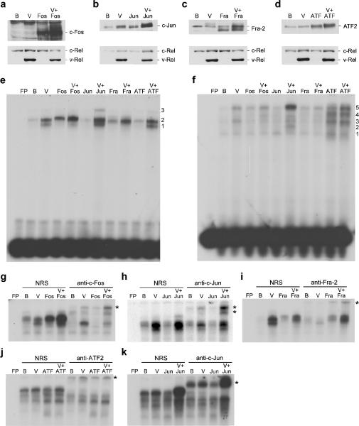
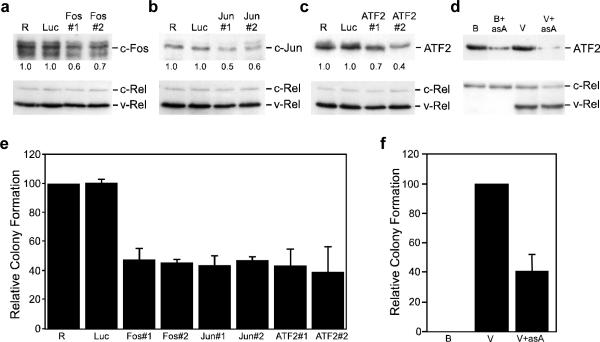
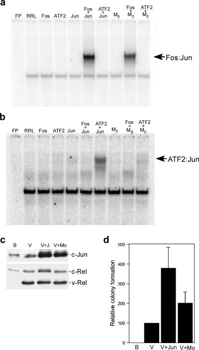
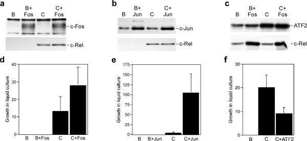
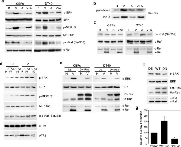
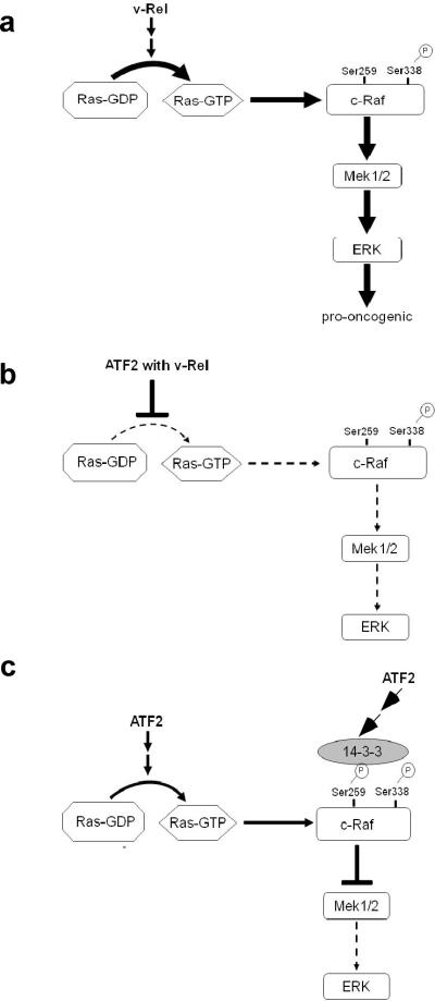
Cell transformation by the v-rel oncogene is mediated by the aberrant expression of genes that are normally tightly regulated by other Rel/NF-kappaB family members. Although a number of genes inappropriately activated or suppressed by v-Rel have been identified, their contributions to the v-Rel transformation process have been poorly characterized. Here, we examine the role of individual AP-1 proteins in v-Rel-mediated transformation. v-Rel-transformed cells exhibit elevated RNA and protein expression of c-Fos, c-Jun and ATF2 and sustained repression of Fra-2. c-Fos and c-Jun are essential in both the initiation and maintenance of v-Rel-mediated transformation, whereas Fra-2 is dispensable. By employing a c-Jun dimerization mutant, we further identified Fos/Jun heterodimers as major contributors to the v-Rel transformation process. The inability of c-Rel to induce the expression of c-Fos and c-Jun contributes to its weaker oncogenic potential relative to v-Rel. Our studies also demonstrate that v-Rel may induce AP-1 members by directly upregulating gene expression (c-fos and ATF2) and by activating pathways that stimulate AP-1 activity. Although elevated expression of ATF2 is also required for v-Rel-mediated transformation, its ectopic overexpression is inhibitory. Investigating the mode of ATF2 regulation revealed a positive feedback mechanism whereby ATF2 induces p38 MAPK phosphorylation to further induce its own activity. In addition, these studies identified Ha-Ras as an effector of v-Rel-mediated transformation and reveal a novel role for ATF2 in the inhibition of the Ras-Raf-MEK-ERK signaling pathway. Overall, these studies reveal distinct and complex roles of AP-1 proteins in Rel/NF-kappaB oncogenesis.


Figures

Figure 1
Figure 1
Expression and DNA binding activity of AP-1 family members expressed from bicistronic retroviruses. DT40 cells were infected with control BIS retroviruses (B), BIS viruses expressing v-Rel (V), c-Fos, c-Jun, Fra-2, and ATF2 alone, or viruses that co-expressed v-Rel with each of the AP-1 family members. Whole cell lysates (25 μg) from infected cells were analyzed by Western blot to evaluate the expression of c-Fos, c-Jun, Fra-2, ATF2, and Rel proteins (panels a–d). Proteins detected by the antisera are indicated on the right of each panel. Nuclear extracts from infected cells were used in EMSAs with [32P]-labeled probes containing AP-1 DNA binding sites specific for Fos:Jun (panel e) or ATF2:Jun heterodimers (panel f). Free probe (FP) and the viruses expressed in the cells that extracts were derived from are indicated above each panel. Numbers to the right of each panel indicate the location of complexes described in the text. Supershift analyses were performed with the extracts described above employing normal rabbit serum (NRS) or antisera specific to c-Fos, c-Jun, Fra-2, and ATF2. Probes containing AP-1 DNA binding sites specific for Fos:Jun (panel g–i) or ATF2:Jun heterodimers (panel j–k) were employed for these studies. The asterisk to the right of each panel indicates the location of the supershifted band(s).
Figure 2
Figure 2
Reduced levels of AP-1 family members inhibit transformation by v-Rel. (a–c) Empty RCAS retroviruses (R) or RCAS viruses expressing an shRNA specific to luciferase (Luc), c-fos (a), c-jun (b), or ATF2 (c) were used to infect 160/2 cells. Whole cell lysates (25–50 μg) from infected cells were harvested ten days after infection and analyzed by Western blot for the expression of c-Fos, c-Jun, ATF2, and Rel proteins. Proteins detected by Western blot are indicated to the right of each panel. The signal corresponding to each AP-1 protein was normalized to that observed in control RCAS infections and values are shown below the top panel. (d) Control BIS retroviruses (B) and BIS viruses expressing v-Rel (V) or antisense ATF2 alone (B+asA), or in combination (V+asA) were used to infect DT40 cells. Whole cell lysates (25 μg) from infected cells were analyzed by Western blot for the expression of ATF2 and Rel proteins. (e) The RCAS-infected 160/2 cells described above were also plated in soft agar and scored for colony formation after eight days. The number of colonies from each experiment was normalized to control RCAS-infected cells. The average and standard deviation of three independent experiments is shown. (f) The BIS viruses described above were used to infect splenic lymphocytes from three-week old chickens. Infected cells were plated in soft agar three days later and colonies scored after ten days. The average and standard deviation of four independent experiments is shown.
Figure 3
Figure 3
Transformation of splenic lymphocytes by viruses co-expressing v-Rel and a c-Jun dimerization mutant. (a and b) DNA binding of the c-Jun dimerization mutant Mo. c-Fos, ATF2, c-Jun, and Mo were in vitro synthesized alone or in combination. Co-translated c-Fos and ATF2 were present in approximately two-fold excess relative to the c-Jun protein synthesized in the reaction. Unprogrammed rabbit reticulocyte lysate (RRL) and in vitro translated proteins were used in EMSAs with [32P]-labeled DNA binding sites specific for Fos:Jun (panel a) and ATF2:Jun (panel b) dimers. (c) Expression of c-Jun proteins in DT40 cells. DT40 cells were infected with control BIS retroviruses (B), BIS viruses expressing v-Rel (V), or viruses that co-expressed v-Rel with c-Jun or Mo. Whole cell lysates (25 μg) from infected cells were analyzed by Western blot to evaluate the expression of Jun and Rel proteins. Proteins detected by the antisera are indicated on the right of each panel. (d) Transformation of cells co-expressing v-Rel and Jun proteins. The viruses described above were used to infect splenic lymphocytes from three-week old chickens. Infected cells were plated in soft agar three days later and colonies scored after ten days. The average and standard deviation of three independent experiments is shown.
Figure 4
Figure 4
Differential effect of enhanced AP-1 expression on the transformation potential of c-Rel. (a–c) Whole cell lysates were prepared from CEFs expressing control BIS retroviruses (B), BIS viruses expressing c-Rel (C), c-Fos, c-Jun, and ATF2 alone, or viruses that co-expressed c-Rel with each of the AP-1 family members. The expression of c-Fos, c-Jun, ATF2, and c-Rel was analyzed by Western blot. Proteins detected by the antisera are indicated on the right of each panel. (d–f) The viruses described above were used to infect splenic lymphocytes from three-week old chickens. Three days later, infected cells were serially diluted in 96-well plates and growth was scored microscopically after ten days. The average and standard deviation of three independent experiments is shown.
Figure 5
Figure 5
Expression of ATF2 alters the activity of MAPKs. (a) Whole cell lysates were prepared from CEFs and DT40 cells expressing control BIS virus (B) and BIS expressing ATF2 (A) and v-Rel alone (V) or in combination (V+A). The expression of ATF2 phosphorylated on Thr69 and Thr71 (p-ATF2), total ATF2, c-Rel, and v-Rel was determined by Western Blot. (b) Whole cell lysates from CEFs and DT40 cells expressing control BIS virus or BIS expressing ATF2 were analyzed by Western blot for the levels of total and active, phosphorylated forms of ERK, JNK, and p38 MAPKs. Expression of ATF2 was also determined. (c) DT40 cells infected with BIS (B) or BIS expressing ATF2 (B+ATF2) were treated for one hour with 10 μM of inhibitors to p38 (SB202190), JNK (SP600125), or MEK1/2 (U0126) kinases, their respective negative controls (SB202474, JNK Inhibitor II Negative Control, U0124), or DMSO alone. Whole cell lysates from treated cells were analyzed by Western blot to determine the levels of phosphorylated and total ATF2. The phosphorylated and total levels of known downstream targets of the p38 (MAPKAP2), JNK (c-Jun), and MEK1/2 (ERK) kinases were analyzed to verify efficacy of the inhibitors.
Figure 6
Figure 6
ATF2 suppresses Ha-Ras activation in v-Rel-expressing cells. (a) Activity of the Raf-MEK-ERK pathway in cells expressing v-Rel. Whole cell lysates were prepared from CEFs and DT40 cells expressing control BIS virus (B) and BIS expressing ATF2 (A) and v-Rel alone (V) or in combination (V+A). The expression of phosphorylated ERK (p-ERK), total ERK, phosphorylated MEK1/2 (p-MEK1/2), total MEK1/2, phosphorylated c-Raf (p-c-Raf Ser338), and total c-Raf was determined by Western Blot analysis. The expression of v-Rel and c-Rel in these lysates was demonstrated in Figure 5a. (b) Regulation of Ha-Ras activity by v-Rel and ATF2. Levels of active Ha-Ras in cell lysates from DT40 cells expressing viruses described above were determined using the Active Ras Pull-Down and Detection Kit (Thermo Scientific). The levels of active (top panel) and total (bottom panel) Ha-Ras in these lysates are shown. (c) Regulation of an inhibitory phosphorylation site on c-Raf by v-Rel and ATF2. Western blot analysis was employed to determine the levels of total and phosphorylated c-Raf (Ser259), c-Rel, and v-Rel in whole cell lysates prepared from the CEFs and DT40 cells described above. (d) Reduction in ATF2 levels increases the activation of the Ras signaling pathway. DT40 cells infected with helper virus CSV (H) or viruses expressing v-Rel (V) were superinfected with control RCAS virus (R) or those expressing two different shRNAs against ATF2 (ATF2#1 and ATF2#2). Whole cell lysates were prepared 10 days post-superinfection and analyzed by Western blot for the levels of total and phosphorylated ERK, MEK1/2, and c-Raf as well as ATF2. (e) Dominant negative HA-Ras inhibits ERK activation in v-Rel-expressing cells. CEFs and DT40 cells infected with helper virus CSV (H) or viruses expressing v-Rel (V) were superinfected with control DS virus or viruses expressing a dominant negative Ha-Ras mutant (DN-Ras). Whole cell lysates were prepared and analyzed by Western blot for the levels of total and phosphorylated ERK, DN-Ras, endogenous HA-Ras, c-Rel, and v-Rel. (f) Ha-Ras regulates ERK activation in a v-Rel transformed cell line. 160/2 cells were infected with control DS virus or viruses expressing wild-type Ha-Ras (WT) or DNRas (DN). Whole cell lysates were prepared and analyzed by Western blot for the levels of total and phosphorylated ERK, ectopic Ras (ect. Ras), endogenous Ha-Ras, c-Rel, and v-Rel. (g) Alterations in Ha-Ras activity affect colony formation of v-Rel transformed cells. 160/2 cells described above were plated in soft agar and scored for colony formation after seven days.
Figure 7
Figure 7
Model for the regulation of Ras-Raf-MEK-ERK pathway by v-Rel and ATF2. (a) v-Rel expression in fibroblast and lymphoid cells results in the induction of Ha-Ras activity, resulting in the phosphorylation and activation of c-Raf, MEK1/2, and ERK. The activation of this pathway by v-Rel plays a key role in transformation by v-Rel. (b) The co-expression of ATF2 with v-Rel blocks v-Rel-mediated activation of the Ras-Raf-MEK-ERK pathway. (c) The ectopic expression of ATF2 alone in DT40 cells induces Ha-Ras activity. However, the activation of the downstream MEK-ERK pathway is blocked by the recruitment of the inhibitory protein 14-3-3 due to the phosphorylation of c-Raf at Ser259.

Similar articles

Cited by

References

    1. Angel P, Hattori K, Smeal T, Karin M. The jun proto-oncogene is positively autoregulated by its product, Jun/AP-1. Cell. 1988;55:875–85. - PubMed
    1. Angel P, Imagawa M, Chiu R, Stein B, Imbra RJ, Rahmsdorf HJ, et al. Phorbol ester-inducible genes contain a common cis element recognized by a TPA-modulated trans-acting factor. Cell. 1987;49:729–39. - PubMed
    1. Bhoumik A, Fichtman B, Derossi C, Breitwieser W, Kluger HM, Davis S, et al. Suppressor role of activating transcription factor 2 (ATF2) in skin cancer. Proc Natl Acad Sci USA. 2008;105:1674–9. - PMC - PubMed
    1. Breitwieser W, Lyons S, Flenniken AM, Ashton G, Bruder G, Willington M, et al. Feedback regulation of p38 activity via ATF2 is essential for survival of embryonic liver cells. Genes Dev. 2007;21:2069–82. - PMC - PubMed
    1. Chaudhary A, King WG, Mattaliano MD, Frost JA, Diaz B, Morrison DK, et al. Phosphatidylinositol 3-kinase regulates Raf1 through Pak phosphorylation of serine 338. Curr Biol. 2000;10:551–4. - PubMed

Publication types

MeSH terms