

Prognostic significance of peritumoral lymphatic vessel density and vascular endothelial growth factor receptor 3 in invasive squamous cell cervical cancer
- PMID: 20563258
- PMCID: PMC2887646
- DOI: 10.1593/tlo.09292
Prognostic significance of peritumoral lymphatic vessel density and vascular endothelial growth factor receptor 3 in invasive squamous cell cervical cancer
Abstract
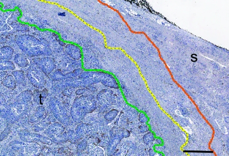
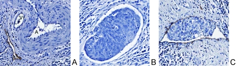
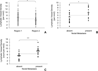
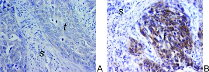
Cervical cancer is known to metastasize primarily by the lymphatic system. Dissemination through lymphatic vessels represents an early step in regional tumor progression, and the presence of lymphatic metastasis is associated with a poor prognosis. In patients who have undergone a radical hysterectomy, lymphovascular space invasion (LVSI), assessed on hematoxylin and eosin-stained slides, is a major factor for adjuvant therapy in patients with cervical cancer. With the advent of a lymphatic endothelial cell-specific marker, such as D2-40, it is now possible to distinguish between blood and lymphatic space invasion (LSI). In this study, the utility of D2-40 was assessed for the detection of lymphatic vessel density (LVD) and identification of LSI. The expressions of vascular endothelial growth factor receptor-3 (VEGFR-3), VEGF-C, tyrosine receptor kinase-2, and angiopoietin-1 were assessed by immunohistochemical methods on 50 patients with squamous cell carcinoma of the cervix. Clinicopathologic characteristics, including pelvic lymph node metastasis, were correlated with the above histochemical findings. We found that lymphangiogenesis, measured by an increase in peritumoral LVD, was significantly associated with positive lymph node status (P < .005). VEGFR-3 expression was significantly associated with LVD (P < .05). D2-40 staining verified LSI (P = .03) and surpassed that of hematoxylin and eosin-identified LVSI (P = .54). In conclusion, lymphangiogenic markers, specifically LVD quantified by D2-40 and VEGFR-3, are independently associated with LSI and lymph node metastasis in patients with early squamous cell carcinoma of the cervix treated with radical hysterectomy and pelvic lymphadenectomy.
Figures
Similar articles
-
Different significance between intratumoral and peritumoral lymphatic vessel density in gastric cancer: a retrospective study of 123 cases.BMC Cancer. 2010 Jun 17;10:299. doi: 10.1186/1471-2407-10-299. BMC Cancer. 2010. PMID: 20565772 Free PMC article.
-
Peritumoral lymphatic vessel density and vascular endothelial growth factor C expression in early-stage squamous cell carcinoma of the uterine cervix.Clin Cancer Res. 2005 Dec 1;11(23):8364-71. doi: 10.1158/1078-0432.CCR-05-1238. Clin Cancer Res. 2005. PMID: 16322297
-
Tumour expression of lymphangiogenic growth factors but not lymphatic vessel density is implicated in human cervical cancer progression.Pathology. 2010 Dec;42(7):629-36. doi: 10.3109/00313025.2010.522174. Pathology. 2010. PMID: 21080871
-
Prognostic Significance of Lymphatic Vessel Density Detected by D2-40 and Its Relation to Claudin-4 Expression in Prostatic Adenocarcinoma.Int J Surg Pathol. 2016 May;24(3):219-26. doi: 10.1177/1066896915611488. Epub 2015 Oct 12. Int J Surg Pathol. 2016. PMID: 26464161
-
Clinical significance of peritumoral lymphatic vessel density and lymphatic vessel invasion detected by D2-40 immunostaining in FIGO Ib1-IIa squamous cell cervical cancer.Cell Tissue Res. 2012 Jun;348(3):515-22. doi: 10.1007/s00441-012-1384-x. Epub 2012 Apr 11. Cell Tissue Res. 2012. PMID: 22492093
Cited by
-
Utility of risk-weighted surgical-pathological factors in early-stage cervical cancer.Br J Cancer. 2013 Apr 2;108(6):1348-57. doi: 10.1038/bjc.2013.78. Epub 2013 Mar 5. Br J Cancer. 2013. PMID: 23462721 Free PMC article.
-
Development of a Deep Learning Model to Identify Lymph Node Metastasis on Magnetic Resonance Imaging in Patients With Cervical Cancer.JAMA Netw Open. 2020 Jul 1;3(7):e2011625. doi: 10.1001/jamanetworkopen.2020.11625. JAMA Netw Open. 2020. PMID: 32706384 Free PMC article.
-
Expression of angiopoietin-2 and vascular endothelial growth factor receptor-3 correlates with lymphangiogenesis and angiogenesis and affects survival of oral squamous cell carcinoma.PLoS One. 2013 Sep 11;8(9):e75388. doi: 10.1371/journal.pone.0075388. eCollection 2013. PLoS One. 2013. PMID: 24040410 Free PMC article.
-
Immunohistochemical study of vascular endothelial growth factor-C/vascular endothelial growth factor receptor-3 expression in oral tongue squamous cell carcinoma: Correlation with the induction of lymphangiogenesis.Oncol Lett. 2015 Oct;10(4):2027-2034. doi: 10.3892/ol.2015.3565. Epub 2015 Aug 4. Oncol Lett. 2015. PMID: 26622791 Free PMC article.
-
Predicting lymph node metastasis in papillary thyroid carcinoma: radiomics using two types of ultrasound elastography.Cancer Imaging. 2025 Feb 13;25(1):13. doi: 10.1186/s40644-025-00832-w. Cancer Imaging. 2025. PMID: 39948651 Free PMC article.
References
-
- Van Trappen PO, Pepper MS. Lymphangiogenesis in human gynaecological cancers. Angiogenesis. 2005;8:137–145. - PubMed
-
- Saharinen P, Tammela T, Karkkainen MJ, Alitalo K. Lymphatic vasculature: development, molecular regulation and role in tumor metastasis and inflammation. Trends Immunol. 2004;25:387–395. - PubMed
-
- Vanan Trappen PO, Pepper MS. Lymphatic dissemination of tumour cells and the formation of micrometastases. Lancet Oncol. 2002;3:44–52. - PubMed
-
- Pepper MS, Tille JC, Nisato R, Skobe M. Lymphangiogenesis and tumor metastasis. Cell Tissue Res. 2003;314:167–177. - PubMed
LinkOut - more resources
Full Text Sources
Miscellaneous