. 2010 Jun 15;8(6):e1000394.
doi: 10.1371/journal.pbio.1000394.

Coupling of a core post-translational pacemaker to a slave transcription/translation feedback loop in a circadian system

Affiliations

Coupling of a core post-translational pacemaker to a slave transcription/translation feedback loop in a circadian system

Ximing Qin et al. PLoS Biol. .

Abstract

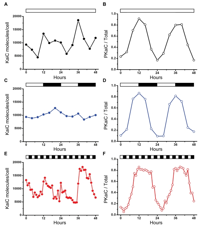
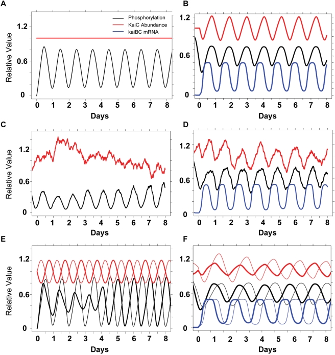
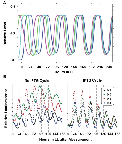
Cyanobacteria are the only model circadian clock system in which a circadian oscillator can be reconstituted in vitro. The underlying circadian mechanism appears to comprise two subcomponents: a post-translational oscillator (PTO) and a transcriptional/translational feedback loop (TTFL). The PTO and TTFL have been hypothesized to operate as dual oscillator systems in cyanobacteria. However, we find that they have a definite hierarchical interdependency-the PTO is the core pacemaker while the TTFL is a slave oscillator that quickly damps when the PTO stops. By analysis of overexpression experiments and mutant clock proteins, we find that the circadian system is dependent upon the PTO and that suppression of the PTO leads to damped TTFL-based oscillations whose temperature compensation is not stable under different metabolic conditions. Mathematical modeling indicates that the experimental data are compatible with a core PTO driving the TTFL; the combined PTO/TTFL system is resilient to noise. Moreover, the modeling indicates a mechanism by which the TTFL can feed into the PTO such that new synthesis of clock proteins can phase-shift or entrain the core PTO pacemaker. This prediction was experimentally tested and confirmed by entraining the in vivo circadian system with cycles of new clock protein synthesis that modulate the phosphorylation status of the clock proteins in the PTO. In cyanobacteria, the PTO is the self-sustained core pacemaker that can operate independently of the TTFL, but the TTFL damps when the phosphorylation status of the PTO is clamped. However, the TTFL can provide entraining input into the PTO. This study is the first to our knowledge to experimentally and theoretically investigate the dynamics of a circadian clock in which a PTO is coupled to a TTFL. These results have important implications for eukaryotic clock systems in that they can explain how a TTFL could appear to be a core circadian clockwork when in fact the true pacemaker is an embedded biochemical oscillator.


Conflict of interest statement

The authors have declared that no competing interests exist.

Figures

Figure 1
Figure 1. In vivo patterns of KaiC abundance and phosphorylation under different illumination conditions.
WT cells (strain AMC149) were assayed in (A, B) LL (constant light; data are averages of three independent experiments), (C, D) LD12:12 (12 h light/12 h dark; data are averages of two independent experiments), (E, F) LD2:2 (2 h light/2 h dark). (A, C, E) KaiC abundance. Based on data of Kitayama and coworkers , the values are normalized to an average number of 10,000 KaiC molecules/cell. (B, D, F) KaiC phosphorylation status, expressed as a ratio of phosphorylated KaiC (P-KaiC) to total KaiC. The KaiC phosphorylation pattern is the most consistent rhythm among the three different in vivo conditions. White/black bars denote light/darkness. Raw data from representative experiments are shown in Figure S1A for KaiC abundance and Figure S1B for KaiC phosphorylation.
Figure 2
Figure 2. Increasing expression of KaiA suppresses the KaiC phosphorylation rhythm and the gene expression rhythm in parallel.
Gene expression was monitored by luminescence from the kaiBCp::luxAB reporter. The expression level of kaiA was under the control of an IPTG-derepressible heterologous trc promoter . (A–G) Inducer at the concentration indicated in the upper left corner of each panel (IPTG, from 0 to 100 µM) was added 18 h before the onset of LL to express KaiA. Each panel depicts the effect of that level of KaiA expression on the luminescence rhythm (measurements for 5 d on duplicate samples—black and red circles) and the KaiC phosphorylation rhythm at peak and trough phases for the first 2 d in LL (immunoblot insets to each panel). (H) Amplitude of the luminescence rhythm as a function of KaiA expression level (driven by varying IPTG concentrations). The amplitude was calculated as the ratio of the first luminescence peak to the first trough using the average of the two replicates (background/trough levels were not subtracted). The data in panels A–H made use of the kaiBCp::luxAB reporter, but similar results were obtained with the psbAIp::luxAB reporter. (I) Amplitude of the phosphorylation rhythm as a function of KaiA expression level/IPTG. The amplitude was calculated as the ratio of the first peak to the first trough and plotted as a function of [IPTG]. Note ordinal scale break. (J) The ratios of the luminescence and phosphorylation rhythms at 0 µM IPTG were set to 1.0, and ratios of the same two rhythms at 100 µM IPTG were set to 0. These normalized amplitude data were then plotted as a function of IPTG concentration.
Figure 3
Figure 3. Cells expressing KaiCEE exhibit damped oscillations in LL that have an abnormally long period and are not temperature compensated under different metabolic conditions.
(A) Cells expressing KaiCWT show robust circadian oscillations in LL at 30°C with a period of ∼25.4 h. (B) Cells expressing KaiCEE (S431E/T432E) exhibit damped, long-period oscillations in LL at 30°C (period ∼58 h, another example is shown in Figure S6). (C) Simulated circadian oscillatory dynamics of KaiC mRNA abundance in the KaiCWT-expressing strain in constant light (LL) in the combined PTO/TTFL model of the KaiABC oscillator. (D) Characteristic simulated oscillatory dynamics of KaiC mRNA in the KaiCEE-expressing strain in constant light (LL) using a generic TTFL without a PTO cycle. In the simulations, KaiB•KaiCEE complexes suppress transcription. A constant light-dependent degradation term (proportional to concentration) removes complexes; translation from mRNA creates KaiB and KaiCEE. The output of simulations from two different parameter sets of the TTFL is shown; damped oscillations of varying period are typical for a wide range of parameters in the model. Details of the model (differential equations and parameter values) can be found in Text S1. (E–H) Rhythms of luminescence in vivo at 24°C (blue traces) versus 30°C (red traces) under different metabolic conditions: (E) WT cultures on agar medium, (F) KaiCEE cultures on agar medium, (G) WT planktonic cultures in liquid medium, (H) KaiCEE planktonic cultures in liquid medium. (I) Period estimates for the data of panels E–H, where WT = KaiCWT and EE = KaiCEE strains. Error bars in panels E–I are S.D. (n values are as follows in panels E–I: 6 for WT at 24°C, 15 for KaiCEE at 24°C, and 5 for both WT and KaiCEE at 30°C). Note that the number of days plotted on the abscissae is different among panels A/B, C/D, and E–H.
Figure 4
Figure 4. Prior entrainment conditions determine the rate of damping in cells expressing KaiCEE.
Cells were in LL at 30°C before and after the following entrainment conditions: (A) one 12 h dark pulse, (B) one 24 h dark pulse, (C) two 12 h dark pulses separated by one 12 h light pulse (i.e., 1.5 cycles of LD12:12), and (D) two 24 h dark pulses separated by one 24 h light pulse (i.e., 1.5 cycles of LD24:24). In all panels, the left ordinate is the luminescence level of the WT strain and the right ordinate is the luminescence level of the KaiCEE strain. In panels A–D, the black traces are the WT luminescence and the red traces are the KaiCEE luminescence (these traces are the average of duplicate measurements, see Figure S8 for all raw data). (E) Damping analysis as the number of days required for the amplitude of the rhythm to decrease to 1/e (≈36.79%) of the starting value. (F) Damping analysis as the number of cycles required for the amplitude of the rhythm to decrease to 1/e. In panels E and F, n = 5 for KaiCWT and n = 7 for each of the KaiCEE sample sets; error bars are SEM.
Figure 5
Figure 5. Simulations derived from the PTO/TTFL model, including resilience and phase-locking.
(A) PTO alone: In the absence of transcription and translation (i.e., for the in vitro reaction or for cells in DD), KaiC abundance is constant (red trace) and simulation of the PTO model indicates a sustained circadian oscillation in KaiC phosphorylation (black trace). Phosphorylation is reported as a fraction of total KaiC. The initial ratios in simulations are 1∶1.5∶1 KaiA:KaiB:KaiC (dimer, tetramer, hexamer) with a nominal initial KaiC concentration of 1 µM (at initial conditions, KaiC is in the unphosphorylated form). (B) Combined PTO/TTFL: Inclusion of a simple TTFL in which hyperphosphorylated KaiB•KaiC complexes that suppress kaiBC transcription show sustained circadian oscillations in KaiC phosphorylation (black), KaiC abundance (red), and kaiBC mRNA (blue) in LL. The phase relationships are consistent with previous in vivo studies of the cyanobacterial system. Simulations are shown following two simulated LD 12:12 cycles. KaiC abundance has been scaled to a mean of 1 (max value ∼3.5 KaiC0). Levels of kaiBC mRNA are reported as a fraction of initially unphosphorylated KaiC ( = KaiC0). (C) PTO resilience as assessed by the effect of noisy unphosphorylated KaiC on the PTO. Random noise in unphosphorylated KaiC was introduced as shown (red trace). KaiC abundance is normalized to a mean of 1.0 (initial concentration is set at 1 µM). The phosphorylation rhythm (black) remains robustly rhythmic despite significant fluctuations in KaiC abundance. (D) Combined PTO/TTFL resilience as assessed by the effect of noisy unphosphorylated KaiC on the PTO/TTFL system. Random noise in unphosphorylated KaiC was introduced at the same level as in Panel C. However, with the inclusion of the TTFL, the same noise fluctuations (as in panel C) result in noisy KaiC abundance oscillations (red trace; KaiC abundance is normalized to a mean of 1.0). The phosphorylation rhythm (black) remains circadian despite significant fluctuations in KaiC but is perturbed by fluctuations in abundance. The abundance of kaiBC mRNA (blue trace) is much less noisy as it reflects the effect of hyper-phosphorylated KaiB•KaiC complexes. Monomer exchange reactions in the PTO decrease the effect of noisy fluctuations in unphosphorylated KaiC fluctuations on the hyper-phosphorylated states. (E) Phase-locking in the PTO. External circadian sinusoidal driving of the abundance of unphosphorylated KaiC in two different phases (0 h, thick lines and 12 h, thin lines) results in the same ultimate asymptotic phase relationship between abundance (red) and phosphorylation (black). At the beginning of the simulation, the phase relationship between KaiC phosphorylation and KaiC abundance is optimal for the thin-trace case and remains so. However, for the thick-trace case, the initial conditions have a non-optimal phase between KaiC phosphorylation and KaiC abundance that resolves into the optimal relationship after about 4 d. (F) Phase-locking in the combined PTO/TTFL. External circadian sinusoidal driving of the abundance of unphosphorylated KaiC as in Panel E in two different phases (0 h, thick lines and 12 h, thin lines) results in the same asymptotic phase relationship between abundance (red), phosphorylation (black), and kaiBC mRNA (blue). The result is similar to that simulated in Panel E except that a final effect on mRNA levels is shown. Details of the models (differential equations and parameter values) can be found in Text S1.
Figure 6
Figure 6. Experimental test of phase locking and entrainment.
(A) Model predictions: simulated phase-locking of kaiBC mRNA rhythms in the combined PTO/TTFL model for four different starting initial phases of mRNA abundance rhythms. Sinusoidal external driving (of unphosphorylated KaiC protein) is implemented as in Figure 5E and 5F. The four phases preferentially lock into a single phase set by the external driving rhythm. The blue trace illustrates the case of initial conditions that are already in the optimal phase relationship to the driving rhythm. (B) Experimental confirmation: cycles of induction of new KaiC synthesis cause phase-locking. Entrainment to four different LD12:12 cycles in which the phase was set to times 0, 6, 12, 18 (i.e., at 6 h intervals) prior to LL release generates four separate populations of cells that are roughly different in phase by 6 h. Cells in four separately phased populations were then treated with two cycles of 0: 0 µM IPTG (i.e. no IPTG cycle, left panel) or 0: 5 µM IPTG (i.e. IPTG cycle: 12 h IPTG, 12 h no IPTG, 12 h IPTG, right panel), and released to free-run in LL. See Figure S9A for an illustration of the experimental protocol.
Figure 7
Figure 7. The core PTO is embedded in a larger TTFL.
The PTO is linked to the damped TTFL (indicated by the pink background circle) by transcription and translation of the kaiABC cluster. Global gene expression is mediated by rhythmic modulation of the activity of all promoters, including those driving the expression of the central clock gene cluster, kaiABC ( = ABC in figure). Rhythmic DNA torsion and/or transcriptional factor activity (e.g., RpaA/SasA) modulate global promoter activities. Cyclic changes in the phosphorylation status of KaiC that mediate the formation of the KaiB•KaiC complex regulate DNA topology/transcriptional factors. The PTO (cycle connected by lavender arrows in upper right quadrant) is determined by KaiC phosphorylation as regulated by interactions with KaiA and KaiB. Robustness is maintained by synchronization of KaiC hexameric status via monomer exchange ,. Monomer exchange is depicted in the figure by “dumbbell” KaiC monomers exchanging with KaiC hexamers in the middle of the PTO cycle; phase-dependent rate of monomer exchange is indicated by the thickness of the double-headed black arrows. The shade of KaiC hexamers (dark versus light blue) denotes conformational changes that roughly equate to kinase versus phosphatase forms. New synthesis of KaiC feeds into the KaiABC oscillator as non-phosphorylated hexamers or as monomers that exchange into pre-existing hexamers. If the new synthesis of KaiC occurs at a phase when hexamers are predominantly hypo-phosphorylated, the oscillation of KaiC phosphorylation is reinforced (enhanced amplitude). If on the other hand, new synthesis of unphosphorylated KaiC happens at a phase when hexamers are predominantly hyper-phosphorylated, this leads to an overall decrease in the KaiC phosphorylation status, thereby altering the phase of the KaiABC oscillator (phase shift) and/or reducing its amplitude.

Similar articles

Cited by

References

    1. Hardin P. E, Hall J. C, Rosbash M. Feedback of the Drosophila period gene product on circadian cycling of its messenger RNA levels. Nature. 1990;343:536–540. - PubMed
    1. Dunlap J. C, Loros J. J, DeCoursey P. J, editors. Chronobiology: biological timekeeping. 2004. Sinauer, Sunderland, MA.
    1. Ishiura M, Kutsuna S, Aoki S, Iwasaki H, Andersson C. R, et al. Expression of a gene cluster kaiABC as a circadian feedback process in cyanobacteria. Science. 1998;281:1519–1523. - PubMed
    1. Xu Y, Mori T, Johnson C. H. Circadian clock-protein expression in cyanobacteria: rhythms and phase setting. EMBO J. 2000;19:3349–3357. - PMC - PubMed
    1. Johnson C. H, Egli M, Stewart P. L. Structural insights into a circadian oscillator. Science. 2008;322:697–701. - PMC - PubMed

Publication types

MeSH terms

Substances