Review
. 2010 Apr;2(4):619-46.
doi: 10.4155/fmc.09.172.

Structural insights into G-quadruplexes: towards new anticancer drugs

Affiliations
Review

Structural insights into G-quadruplexes: towards new anticancer drugs

Danzhou Yang et al. Future Med Chem. 2010 Apr.

Abstract

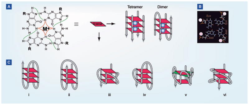
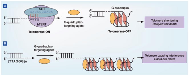
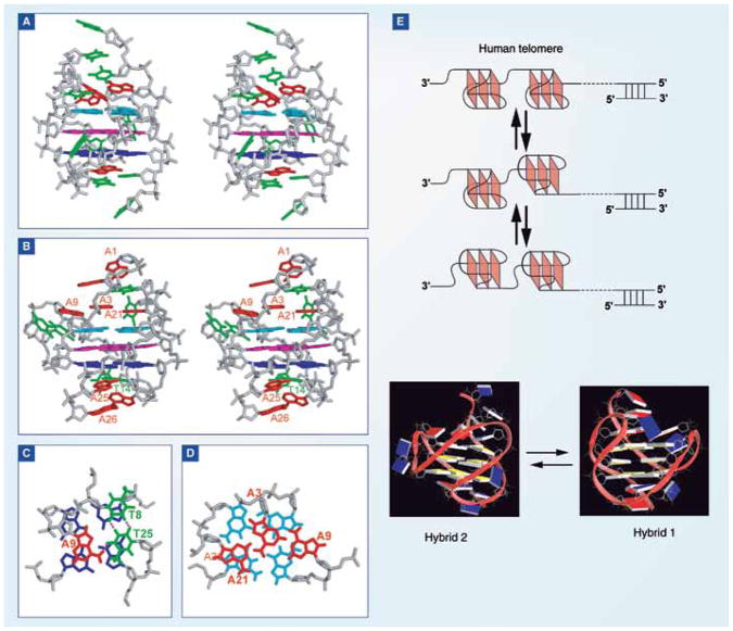
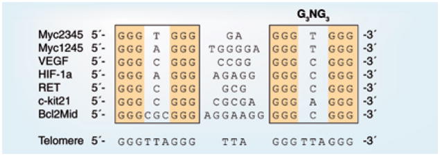
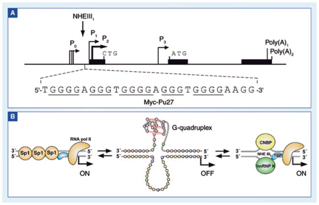
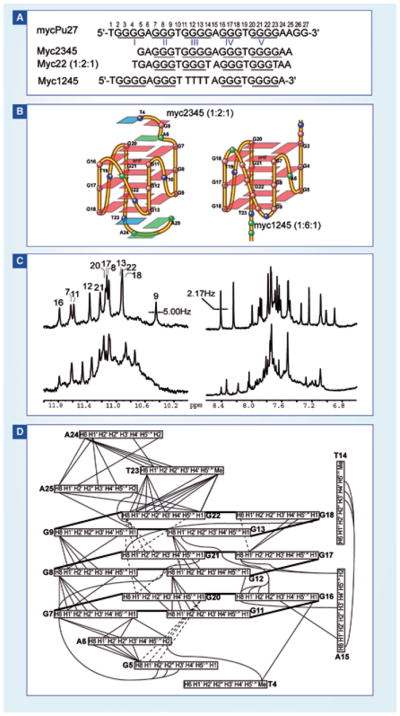
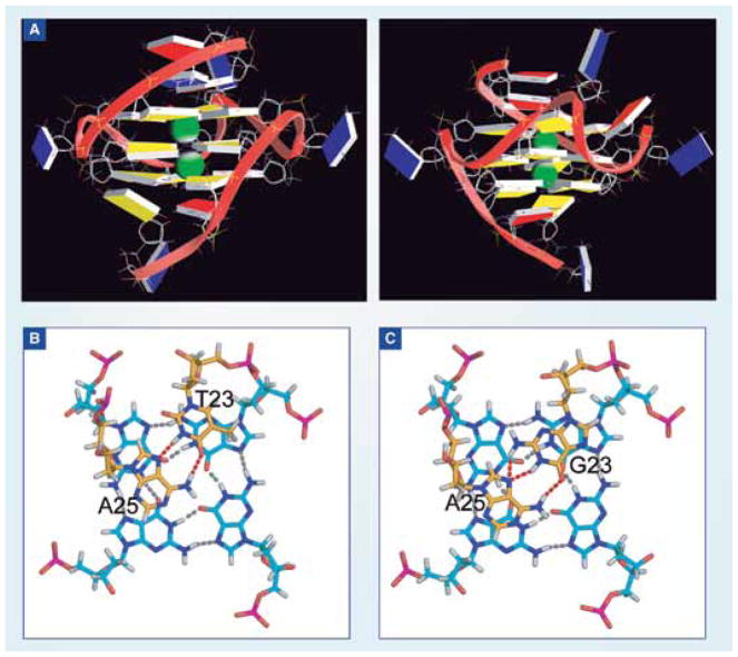
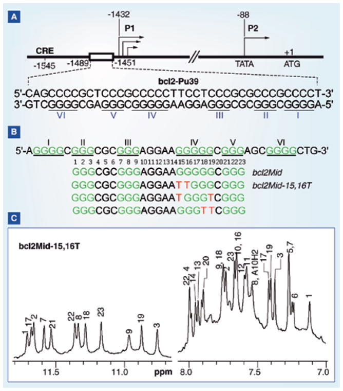
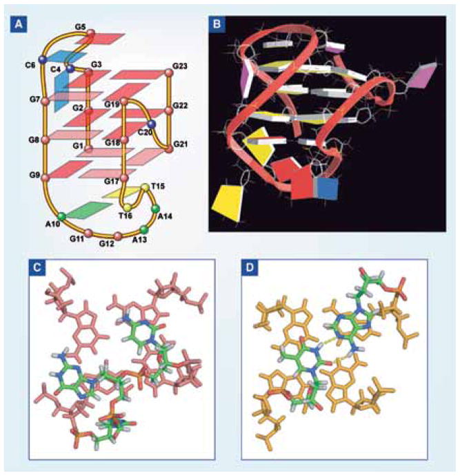
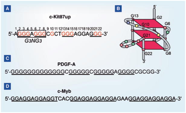
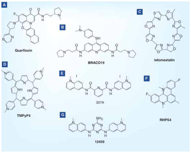
DNA G-quadruplexes are DNA secondary structures formed in specific G-rich sequences. DNA sequences that can form G-quadruplexes have been found in regions with biological significance, such as human telomeres and oncogene-promoter regions. DNA G-quadruplexes have recently emerged as a new class of novel molecular targets for anticancer drugs. Recent progress on structural studies of the biologically relevant G-quadruplexes formed in human telomeres and in the promoter regions of human oncogenes will be discussed, as well as recent advances in the design and development of G-quadruplex-interactive drugs. DNA G-quadruplexes can readily form in solution under physiological conditions and are globularly folded nucleic acid structures. The molecular structures of intramolecular G-quadruplexes appear to differ from one another and, therefore, in principle may be differentially regulated and targeted by different proteins and drugs.


Figures

Figure 1
Figure 1
(A) G-tetrad, a square-planar alignment of four guanines connected by cyclic Hoogsteen hydrogen bonding between the N1, N2 and O6, N7 of guanine bases (left). The H1–H1 and H1–H8 connectivity patterns detectable in NOESY experiments is also shown. A schematic tetrameric and dimeric G-quadruplex composed of three G-tetrads (right). Cations (K+ or Na+), shown as blue balls, are needed to stabilize G-quadruplexes by coordinating with the eight electronegative carbonyl oxygen O6 atoms of the adjacent G-tetrads. (B) The guanines in a G-tetrad can adopt either syn or anti glycosidic conformation. The guanines from the parallel G-strands adopt the same glycosidic conformation and the guanines from the antiparallel G-strands adopt the opposite glycosidic conformations. (C) Examples of monomeric (intramolecular) G-quadruplexes with different folding structures.
Figure 2
Figure 2
(A) Mechanism of telomerase inhibition by G-quadruplex-targeting compounds. (B) Mechanism of drug-mediated interference of telomere capping by G-quadruplex-targeting compounds.
Figure 3
Figure 3
(A) Four-G-tract human telomeric sequences with different flanking sequences. The numbering system is shown above wtTel27. The major conformation formed in each sequence is indicated. (B) The imino proton region of the 1D 1H NMR of (i) wtTel22, (ii) Tel26 with assignment and (iii) wtTel26 with assignment in K+ solution. (C) Titration experiments of K+ in the presence of 150 mM Na+ for Tel26 (left), and titration experiments of Na+ in the presence of 100 mM K+ for Tel26 (right), monitored by circular dichroism spectroscopy.
Figure 4
Figure 4
(A) Folding topology of the basket-type intramolecular G-quadruplex formed by wtTel22 in Na+ solution as determined by NMR (i). Folding topology of the propeller-type parallel-stranded intramolecular G-quadruplex formed by wtTel22 in the presence of K+ in crystalline state (ii). Folding topologies of the hybrid-1 (major conformation in Tel26) (iii, left) and hybrid-2 (major conformation in wtTel26) (iii, right) intramolecular G-quadruplexes in K+ solution. The numbering system is based on wtTel26. Yellow box: (anti) guanine; red box: (syn) guanine. (B) A model of the interconversion between the basket-type (Na+) and the hybrid-type (K+) telomeric G-quadruplexes through a strand-reorientation mechanism. A two-tetrad form is likely to be a transition intermediate of the interconversion between different telomeric structures.
Figure 5
Figure 5
(A) Stereo view of the representative NMR structure of the hybrid-2 telomeric G-quadruplex formed by wtTel26 in K+ solution. (B) Stereo view of the representative NMR structure of the hybrid-1 telomeric G-quadruplex formed by Tel26 in K+ solution. (C) The bottom view of the T:A:T triple capping the bottom G-tetrad (blue) of hybrid-2 telomeric G-quadruplex, with the potential hydrogen bonds shown as dashed lines. (D) Top view of the adenine triple (red) capping the top G-tetrad (cyan) of hybrid-1 telomeric G-quadruplex. The top (from 5′-end) G-tetrad is in cyan, the middle G-tetrad is in magenta, and the bottom G-tetrad is in blue. (E) A model showing a DNA secondary structure composed of compact-stacking multimers of hybrid-type G-quadruplexes in human telomeres, with an equilibrium between hybrid-1 and hybrid-2 forms in K+ solution. The representative NMR structures, which are distinct, of hybrid-1 and hybrid-2 telomeric G-quadruplexes are shown (guanine: yellow; adenine: red; thymine: blue).
Figure 6
Figure 6
Comparison of G-quadruplex-forming sequences in selected gene promoters. The telomeric sequence is also shown as a comparison. All the promoter G-rich sequences shown contain the G3NG3 motif; except for the BCL-2 sequence, they have all been shown to form parallel-stranded G-quadruplexes.
Figure 7
Figure 7
(A) The promoter structure of the human c-MYC gene. The G-rich NHE III1 is shown, with guanine runs underlined. (B) Alternative forms of the NHE III1 of the c-MYC promoter associated with transcriptional activation or silencing.
Figure 8
Figure 8
(A) The c-MYC promoter sequence and its modifications. (B) The folding structure of the major G-quadruplex formed in the c-MYC promoter, Myc2345(1:2:1) (left) and the minor G-quadruplex, Myc1245 (right). (C) 1D NMR spectra of the T-Myc2345 sequence (bottom) and its major loop isomer Myc22 (top). (D) Inter-residue NOEs of MYC22, which forms the major G-quadruplex of the c-MYC promoter. The NOE connectivities clearly define the quadruplex conformation and provide distance restraints for structure calculation.
Figure 9
Figure 9
(A) Representative NMR structure of the major c-MYC promoter G-quadruplex formed by Myc22, a parallel-stranded structure, in two opposite views. Two potassium ions coordinated between the G-tetrads are included for the calculation and are shown as green spheres. (Guanine: yellow; adenine: red; thymine: blue) The 3′-end view of the G-quadruplex with mutant T23 (B) and wild-type G23 (C). The hydrogen bonds of the top tetrad (black) and the T/G23:A25 base pair (green) are shown in dashed lines.
Figure 10
Figure 10
(A) Promoter structure of the human BCL-2 gene. The G/C-rich region of the promoter is shown, with guanine runs underlined. (B) The BCL-2 promoter sequence and its modifications. (C) 1D NMR spectrum of the bcl2Mid-15,16T, with assignments, which forms the major G-quadruplex in the BCL-2 promoter.
Figure 11
Figure 11
(A) Folding structure of the major G-quadruplex formed in the BCL-2 promoter. (B) A representative NMR structure of the major BCL-2 promoter G-quadruplex. The (C) 5′-end and (D) 3′-end view of the major BCL-2 promoter G-quadruplex. The hydrogen bonds of the A10:T15 base pair are shown in dashed lines.
Figure 12
Figure 12
(A) Sequence of one G-rich region of the human c-KIT gene promoter (c-KIT87up). (B) The folding topology of the NMR-determined structure formed by c-KIT87up [168]. (C) The sequence of the G-rich region of the human PDGF-A gene promoter. (D) The sequence of the G-rich region of the human c-MYB gene promoter.
Figure 13
Figure 13
G-quadruplex-interactive small-molecule compounds.

Similar articles

Cited by

References

    1. Gellert M, Lipsett MN, Davies DR. Helix formation by guanylic acid. Proc Natl Acad Sci USA. 1962;48:2013–2018. - PMC - PubMed
    1. Sen D, Gilbert W. A sodium-potassium switch in the formation of four-stranded G4-DNA. 1990;344(6265):410–414. - PubMed
    1. Hud NV, Plavec J. The role of cations in determining quadruplex structure and stability. In: Neidle S, editor. Quadruplex Nucleic Acids. Royal Society of Chemistry Publishing; Cambridge UK: 2006. pp. 100–130.
    1. Neidle S, Parkinson G. Telomere maintenance as a target for anticancer drug discovery. Nat Rev Drug Discov. 2002;1(5):383–393. - PubMed
    1. Dai JX, Carver M, Yang DZ. Polymorphism of human telomeric quadruplex structures. Biochimie. 2008;90(8):1172–1183. - PMC - PubMed

Publication types

MeSH terms