. 2010 Oct;62(10):2984-94.
doi: 10.1002/art.27614.

Impaired activation-induced cell death promotes spontaneous arthritis in antigen (cartilage proteoglycan)-specific T cell receptor-transgenic mice

Affiliations

Impaired activation-induced cell death promotes spontaneous arthritis in antigen (cartilage proteoglycan)-specific T cell receptor-transgenic mice

Ferenc Boldizsar et al. Arthritis Rheum. 2010 Oct.

Abstract

Objective: To investigate whether genetic preponderance of a T cell receptor (TCR) recognizing an arthritogenic peptide of human cartilage proteoglycan (PG) is sufficient for development of arthritis.

Methods: We performed a longitudinal study using BALB/c mice expressing a TCR that recognizes the arthritogenic ATEGRVRVNSAYQDK peptide of human cartilage PG. PG-specific TCR-transgenic (PG-TCR-Tg) mice were inspected weekly for peripheral arthritis until 12 months of age. Peripheral joints were examined histologically, and T cell responses, T cell activation markers, serum cytokines, and autoantibodies were measured. Apoptosis and signaling studies were performed in vitro on T cells from aged PG-TCR-Tg mice.

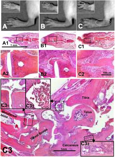
Results: Spontaneous arthritis developed as early as 5-6 months of age, and the incidence increased to 40-50% by 12 months of age. Progressive inflammation began with cartilage and bone erosions in the interphalangeal joints, and later expanded to the proximal joints of the front and hind paws. Spontaneous arthritis was associated with a high proportion of activated CD4+ T cells, enhanced interferon-γ and interleukin-17 (IL-17) production, and elevated levels of serum autoantibodies. PG-TCR-Tg mice lacking IL-4 developed arthritis earlier and at a higher incidence than IL-4-sufficient mice. Antigen-specific activation-induced cell death was diminished in vitro in CD4+ T cells of PG-TCR-Tg mice with spontaneous arthritis, especially in those lacking IL-4.

Conclusion: The presence of CD4+ T cells expressing a TCR specific for an arthritogenic PG epitope is sufficient to trigger spontaneous autoimmune inflammation in the joints of BALB/c mice. IL-4 appears to be a negative regulator of this disease, through attenuation of activation-induced cell death.


Figures

Figure 1
Figure 1
Timeline of the development of spontaneous arthritis in the 5/4E8 peptide (ATEGRVRVNSAYQDK)-specific PG-TCR-Tg (PG-TCR-Tg) mice. Swelling and mild redness of the distal interphalangeal joints were considered the first signs of inflammation. Values are calculated as percent of arthritic animals per total number of PG-TCR-Tg mice in each group followed up to 12 months of age. (A) Spontaneous arthritis in 28 female 5/4E8 peptide-specific PG-TCR-Tg BALB/c mice. (B) Spontaneous arthritis in 20 female IL-4-deficient PG-TCR-Tg (PG-TCR-Tg/IL-4−/−) BALB/c mice.
Figure 2
Figure 2
Macroscopic and histology images of front and hind paws of (A) wild type (healthy 9-month-old BALB/c), (B) spontaneously arthritic PG-TCR-Tg/IL-4+/+ and (C) PG-TCR-Tg/IL-4−/− mice. Corresponding sections (at low and high magnifications) of fingers underneath the macroscopic images are depicted in panels A1–A2, B1–B2 and C1–C2. Thickening of the distal interphalangeal joints and phalanges and loss of nails were the earliest macroscopic abnormalities, which were followed by progression of inflammation to the proximal interphalangeal, metatarsophalangeal and tarso-matatarsal or carpo-metacarpal joints. Panel C3 is a low magnification montage picture of a hind paw (ankle area) from a 1-year-old PG-TCR-Tg/IL-4−/− BALB/c mouse (approximately 6 months after arthritis onset). Extensive cartilage and bone erosions of affected joints are the prominent histopathological abnormalities demonstrated in high-magnification inserts (C31 and C33). Dominantly mononuclear cells infiltrate (C32) the synovium and the joint cavities. Contours of multinuclear osteoclasts are indicated by white broken lines and arrowheads (C33). Representative hematoxylin-stained sections are shown at 4× (A1, B1, C1 and C3), 10× (A2, B2 and C2) and 40× (C31, C32 and C33) original magnification.
Figure 3
Figure 3
Antigen-specific activation-induced apoptosis in PG-TCR-Tg CD4+ T cells. In vitro 5/4E8 peptide stimulation-induced apoptosis of purified CD4+ T cells of spleens (from IL-4-deficient and IL-4-sufficient PG-TCR-Tg BALB/c mice) were compared. (A) Percent of live (Annexin VPI), early apoptotic (Annexin V+PI) and late apoptotic (Annexin V+PI+) cells. Bars represent mean ± SEM values calculated from the data of three mice in each group on days two and three. Significant (p<0.05) differences are indicated by asterisks. Representative flow cytometric contour plots show the distribution of (B) PG-TCR-Tg /IL-4+/+ and (C) PG-TCR-Tg/IL-4−/− T cells according to their Annexin V and PI staining after three days culture in the presence of TCR-specific 5/4E8 peptide presented by semi-confluent irradiated A20 cells. Numbers in the quadrants of contour plots show the percent of total cells.
Figure 4
Figure 4
Phosphorylation changes upon TCR stimulation in PG-TCR-Tg T cells. Flow cytometric comparison of the in vitro 5/4E8 peptide-induced signaling was performed using purified T cells (more than 95% CD4+Vβ4+) harvested from spleens of old (≥9 months) PG-TCR-Tg/IL-4+/+ or PG-TCR-Tg/IL-4−/− mice. (A and B) Diagrams show values of mean fluorescence intensities (MFI) from four representative samples, measured in the FL2 channel after labeling the cells (A) with anti-phospho-Zap70-PE or (B) anti-phospho-ERK1/2-PE antibodies. (C) Representative FL-2 histogram plots show the Zap70 phosphorylation measured at different time points in the CD4+ T cells of PG-TCR-Tg /IL-4+/+ and PG-TCR-Tg/IL-4−/− mice. (D) Representative FL-2 histogram plots show the ERK1/2 phosphorylation measured at the indicated time points in the CD4+ T cells of PG-TCR-Tg/IL-4+/+ and PG-TCR-Tg/IL-4−/− mice. Numbers inside the panels indicate the MFI value, representing the phosphorylation status of Zap70 or ERK1/2. Time points are indicated between the histogram plots of panels C and D. The vertical black line in the histogram plots (C, D) is set at the MFI value of the control sample.

References

    1. Campbell IK, Kinkel SA, Drake SF, van Nieuwenhuijze A, Hubert FX, Tarlinton DM, et al. Autoimmune regulator controls T cell help for pathogenetic autoantibody production in collagen-induced arthritis. Arthritis Rheum. 2009;60:1683–93. - PubMed
    1. Trentham DE, Dynesius RA, Rocklin RE, David JR. Cellular sensitivity to collagen in rheumatoid arthritis. N Engl J Med. 1978;299:327–32. - PubMed
    1. Glant T, Csongor J, Szücs T. Immunopathologic role of proteoglycan antigens in rheumatoid joint diseases. Scand J Immunol. 1980;11:247–52. - PubMed
    1. Courtenay JS, Dallman MJ, Dayan AD, Martin A, Mosedale B. Immunization against heterologous type II collagen induces arthritis in mice. Nature. 1980;282:666–8. - PubMed
    1. Glant TT, Mikecz K, Arzoumanian A, Poole AR. Proteoglycan-induced arthritis in BALB/c mice. Clinical features and histopathology. Arthritis Rheum. 1987;30:201–12. - PubMed

Publication types