. 2010 Jul 27;49(29):6263-75.
doi: 10.1021/bi100517x.

Mechanisms of 4-hydroxy-2-nonenal induced pro- and anti-apoptotic signaling

Affiliations

Mechanisms of 4-hydroxy-2-nonenal induced pro- and anti-apoptotic signaling

Pankaj Chaudhary et al. Biochemistry. .

Abstract

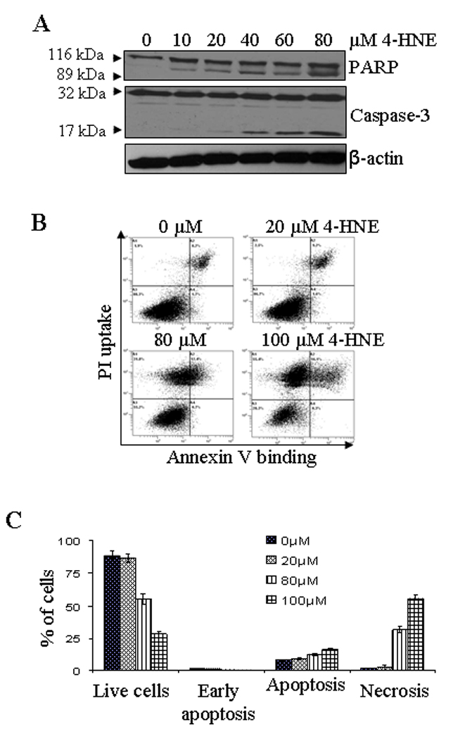
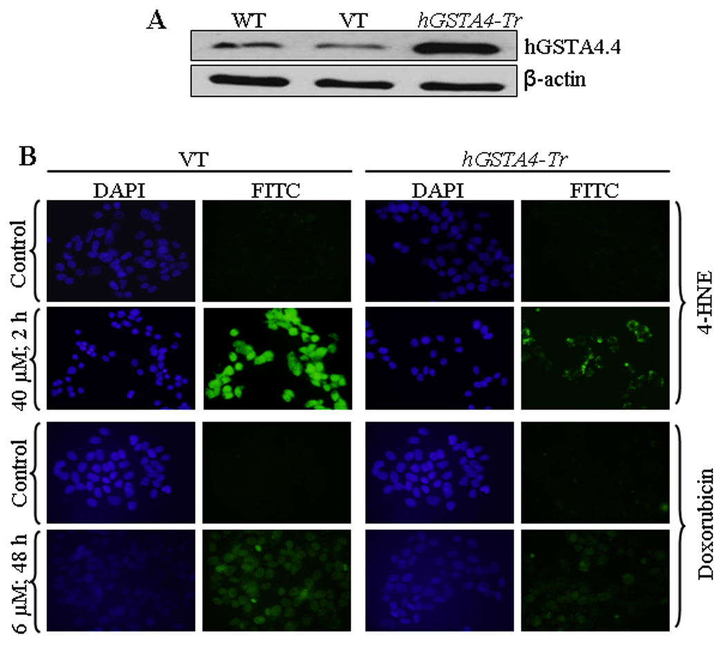
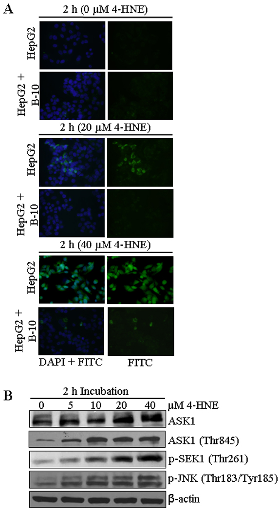
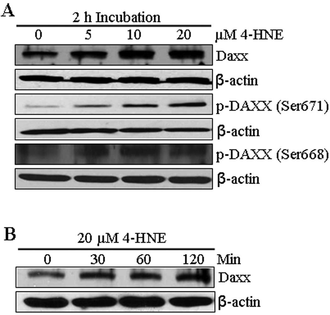
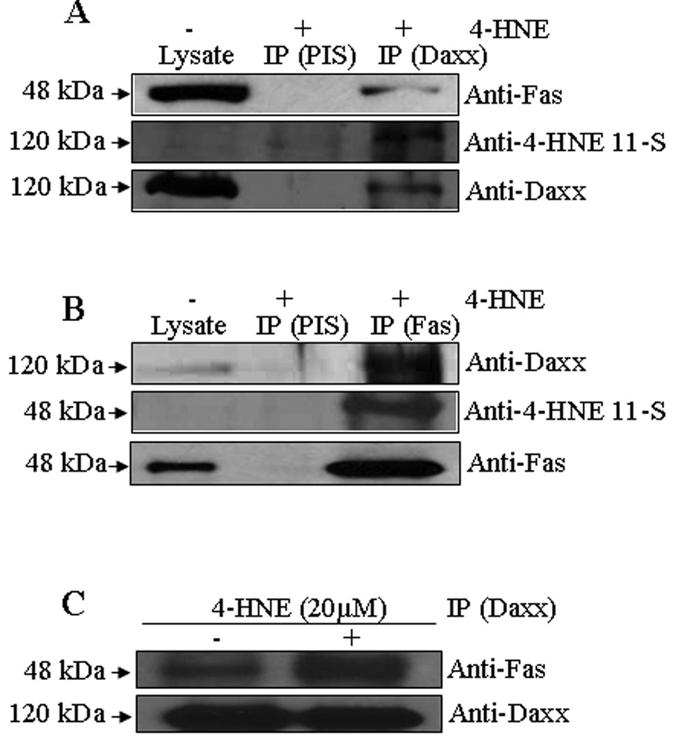
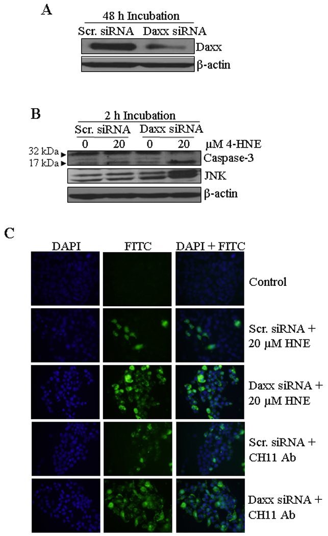
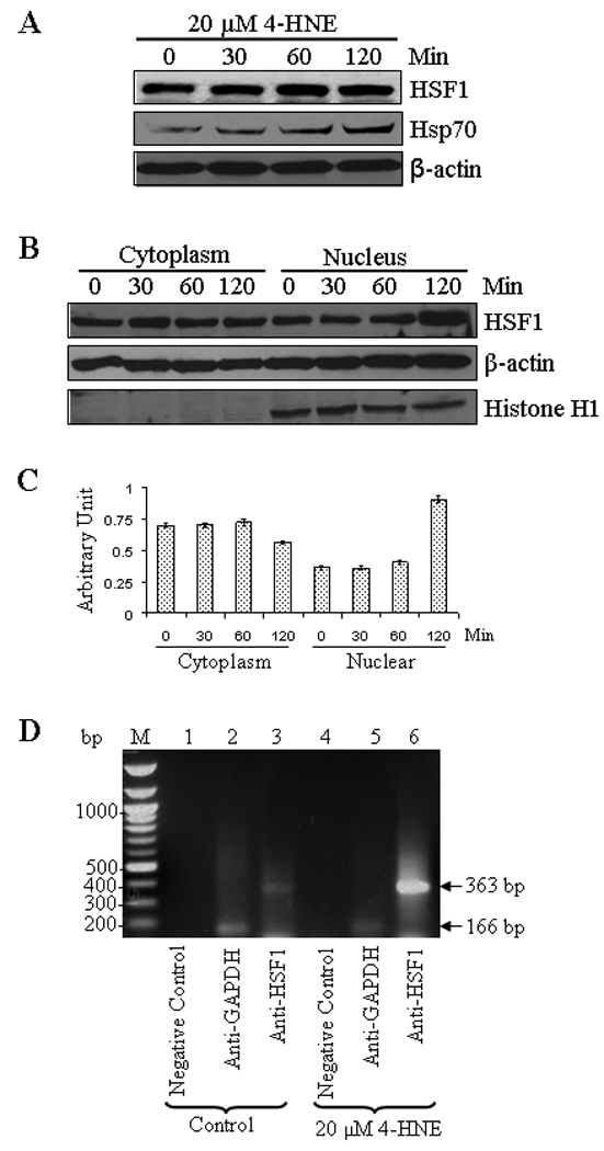
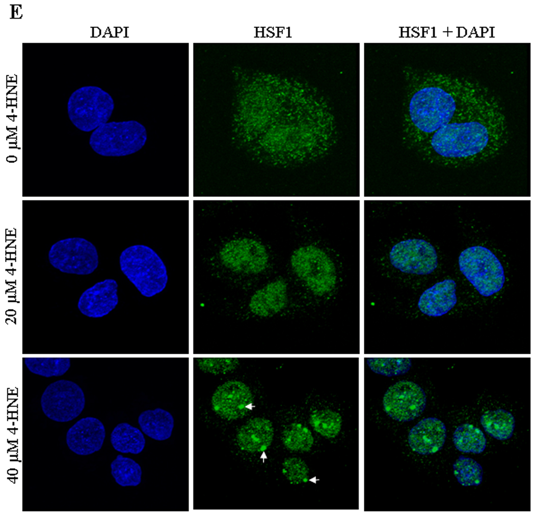
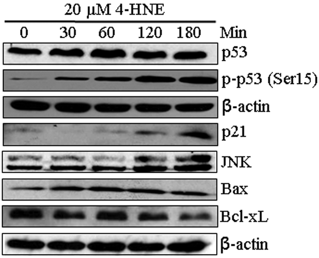
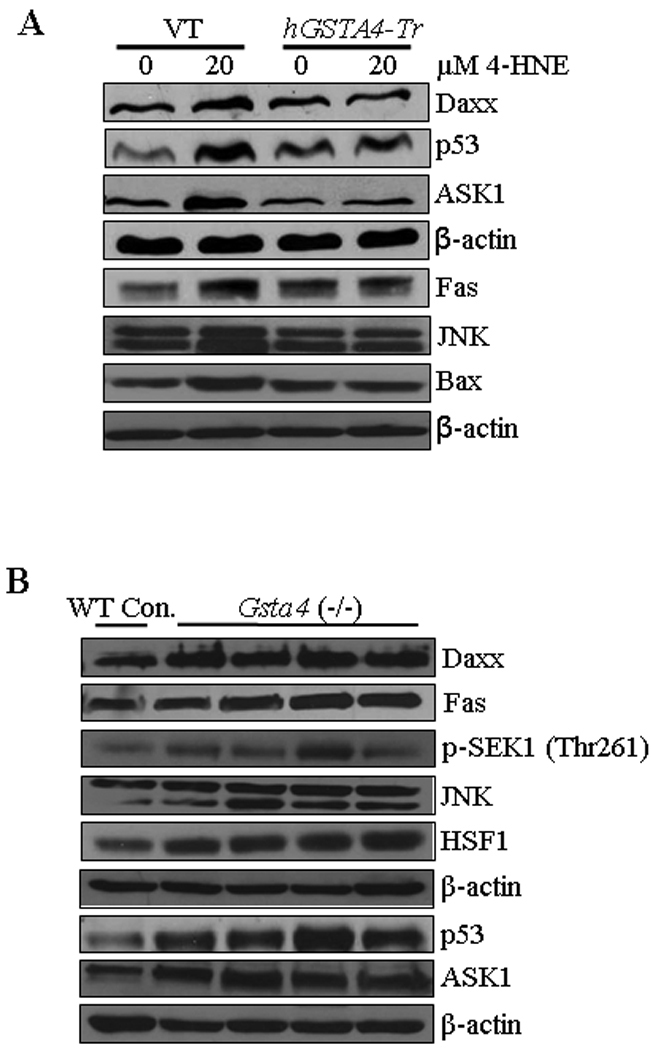
In recent years, 4-hydroxy-2-nonenal (4-HNE) has emerged as an important second messenger in cell cycle signaling. Here, we demonstrate that 4-HNE induces signaling for apoptosis via both the Fas-mediated extrinsic and the p53-mediated intrinsic pathways in HepG2 cells. 4-HNE induces a Fas-mediated DISC independent apoptosis pathway by activating ASK1, JNK, and caspase-3. Parallel treatment of 4-HNE to HepG2 cells also induces apoptosis by the p53 pathway through activation of Bax, p21, JNK, and caspase-3. Exposure of HepG2 cells to 4-HNE leads to the activation of both Fas and Daxx, promotes the export of Daxx from the nucleus to cytoplasm, and facilitates Fas-Daxx binding. Depletion of Daxx by siRNA results in the potentiation of apoptosis, indicating that Fas-Daxx binding in fact is inhibitory to Fas-mediated apoptosis in cells. 4-HNE-induced translocation of Daxx is also accompanied by the activation and nuclear accumulation of HSF1 and up-regulation of heat shock protein Hsp70. All these effects of 4-HNE in cells can be attenuated by ectopic expression of hGSTA4-4, the isozyme of glutathione S-transferase with high activity for 4-HNE. Through immunoprecipitation and liquid chromatography-tandem mass spectrometry, we have demonstrated the covalent binding of 4-HNE to Daxx. We also demonstrate that 4-HNE modification induces phosphorylation of Daxx at Ser668 and Ser671 to facilitate its cytoplasmic export. These results indicate that while 4-HNE exhibits toxicity through several mechanisms, in parallel it evokes signaling for defense mechanisms to self-regulate its toxicity and can simultaneously affect multiple signaling pathways through its interactions with membrane receptors and transcription factors/repressors.


Figures

Figure 1
Figure 1. Effect of 4-HNE on HepG2 cells
(A) Western blot analysis showing the caspase-3 and PARP cleavage: HepG2 cells were treated with different concentrations of 4-HNE (0–80 µM) for 2 h at 37°C. The cells were scraped, collected and then washed with ice cold PBS, and the cell lysates were prepared as described in Materials and methods section. The cells extracts (50 µg of protein) were subjected to Western blot analyses using anti-caspase-3 and anti-PARP antibodies. Anti-β-actin antibody was used as loading control. (B) Flow cytometry analysis of cells treated with 4-HNE: HepG2 cells (4 × 10⁵) were grown and treated with different concentrations of 4-HNE for 2 h at 37°C in full serum medium. After treatment, the cells were harvested by trypsinization and washed with ice cold PBS and resuspended in 400 µL of cold Annexin binding buffer containing 5 µL of Annexin V-FITC and 5 µL of 0.1 mg/mL propidium iodide. The cells were incubated at room temperature for 10 min. in the dark and were analyzed by flow cytometry using a Beckman Coulter Cytomics FC500 flow cytometer. (C) Bar graph showing the percent of cells present in apoptosis vs necrosis of the panel B.
Figure 2
Figure 2. Effect of hGSTA4 transfection on 4-HNE and doxorubicin-induced apoptosis
(A) Western blot analysis shows the expression of hGSTA4-4 in wild type, empty vector- (VT) and hGSTA4 transfected HepG2 cells. (B) In situ analysis of activated caspase-3 in VT and hGSTA4 transfected HepG2 cells. The cells (2 × 10⁴) were treated with either 40 µM 4-HNE for 2 h or 6 µM doxorubicin for 48 h. The activation of caspase-3 in these cells was examined by staining with 10 µM CaspACE™ FITC-VAD-FMK in situ marker according to the manufacturer’s instructions. The slides were mounted with Vectashield DAPI mounting medium and observed with a fluorescence microscope (Olympus) using the standard filter sets for DAPI and FITC.
Figure 3
Figure 3. Effect of 4-HNE on Fas mediated apoptosis in HepG2 cells
(A) In situ detection of Fas-mediated apoptosis in HepG2 cells. 2 × 10⁴ cells were grown on glass cover slides and after pretreatment with or without antagonistic anti-Fas (B-10) antibodies for 2 h (250 ng/mL) HepG2 cells were treated with 0, 20 and 40 µM 4-HNE for 2 h followed by the addition of in situ caspase marker and fixation. The slides were mounted with Vectashield DAPI mounting medium and observed under a fluorescence microscope (Olympus). The photographs were taken at 400× magnification. (B) The cells were treated with 0, 5, 10, 20 and 40 µM 4-HNE for 2 h at 37°C. Total protein lysates (30 µg) were analyzed by western blotting for ASK1, p-ASK1 (Thr845), p-SEK1 (Thr261), and p-JNK (Thr183/Tyr185). β-actin was used as a loading control.
Figure 4
Figure 4. 4-HNE induced expression of Daxx and phospho-Daxx in HepG2 cells
(A) The cells were treated with 0, 5, 10, and 20 µM 4-HNE for 2 h at 37°C. Total protein lysates (30 µg) were analyzed by Western blotting for the expression of Daxx, p-Daxx (Ser671) and p-Daxx (Ser668). β-actin was used as a loading control. (B) The cells were treated with 20 µM 4-HNE for 0, 30, 60, 120 min at 37°C. Total protein lysates were subjected to western blot analyses for Daxx expression. β-actin was used as a loading control.
Figure 5
Figure 5. Effect of 4-HNE induced cytoplasmic translocation of nuclear Daxx in HepG2 cells
(A) The cells (4 × 10⁵) were grown and treated with 20 µM 4-HNE for 0, 1 and 2 h, separately, at 37°C. After completion of incubation, cells were scraped, collected and washed with ice cold PBS. Cytoplasmic and nuclear extracts of the pelleted cells were prepared using Imgenex kit as per manufacturer’s instructions. The extracts (50 µg of protein) were subjected to western blot analysis for the detection of Daxx, β-actin and Histone H3. Antibodies against β-actin and Histone H3 were used to determine the purity of the cytoplasmic and nuclear fractions respectively. (B) Bar chart showing the densitometric analysis of Daxx bands of immunoblot of panel A. (C) Confocal immunofluorescence analysis of nuclear Daxx translocation. HepG2 cells were grown on glass cover slips and untreated and treated (20 µM 4-HNE for 2 h) cells were fixed, permeabilized and incubated with polyclonal anti-Daxx antibody, followed by fluorescein (FITC) conjugated secondary antibody. DAPI staining shows the nucleus. Slides were analyzed using Zeiss LSM 510META laser scanning fluorescence microscope.
Figure 6
Figure 6. Co-immunoprecipitation of Fas and Daxx
Total protein lysates were collected from control and 4-HNE-treated (20 µM for 2 h) HepG2 cells. The cell lysates were immunoprecipitated with anti-Fas and anti-Daxx antibodies as described in the materials and methods followed by Western blotting to check the expression of different proteins as indicated in the panel. Immunoprecipitation with pre-immune serum (PIS) was used as control. (A) The immunoprecipitate of Daxx (IP Daxx) were probed with antibodies indicated in the panel shows the presence of Fas, 4-HNE-Daxx adduct and Daxx. (B) The immunoprecipitate of Fas (IP Fas) was probed with antibodies indicated in the panel and shows the presence of Daxx and 4-HNE-Fas adduct. (C) The cell lysates were immunoprecipitated with anti-Daxx antibodies from control and 4-HNE-treated (20 µM for 2 h) HepG2 cells followed by Western blotting to check the expression of Fas and Daxx proteins as indicated in the panel.
Figure 7
Figure 7. Silencing of Daxx expression in HepG2 cells by siRNA and potentiation of apoptosis by 4-HNE and CH11 antibody
(A) Silencing of Daxx was performed by the ON-TARGET plus SMART pool Daxx siRNA as per the manufacturer’s instructions (Thermo Scientific Dharmacon) and control cells were treated with ON-TARGET plus Non-targeting siRNA in a similar way. After transfection, the cells were harvested after 48 h and the expression level of Daxx was examined by Western blot analysis. (B) Western blot analysis showing the enhanced activation of caspase-3 and JNK in Daxx depleted cells after 4-HNE treatment (20 µM HNE for 2 h). (C) Enhanced activation of apoptosis was also performed by In Situ immunofluorescence study. The cells (2 × 10⁵) were grown on glass cover slips in twelve-well plate and transfected with non targeting siRNA or Daxx siRNA. After 48 h of siRNA transfection, the cells were treated with either 50 µg/well Fas-agonistic antibodies (CH11) or 20 µM 4-HNE for 2 h at 37°C. The activation of caspase-3 in these cells was examined by CaspACE FITC-VAD-FMK in situ marker as per the manufacturer’s instructions and then observed under a fluorescence microscope.
Figure 8
Figure 8. 4-HNE induced expression of heat shock factor 1 and heat shock protein Hsp70 in HepG2 cells
(A) The cells were treated with 20 µM 4-HNE for different time points at 37°C. Total protein lysates were analyzed by western blotting for HSF1, and Hsp70. β-actin was used as a loading control. (B) Effect of 4-HNE induced nuclear translocation of HSF1 in HepG2 cells was analyzed by Western blot analysis. The cells (4 × 10⁵) were grown and treated with 20 µM 4-HNE for 0, 30, 60 and 120 min, separately, at 37°C. After 4-HNE incubation, cells were scraped, collected and washed with ice cold PBS. Cytoplasmic and nuclear extracts of the pelleted cells were prepared according to the Imgenex kit. The extracts (30 µg of protein) were subjected to western blot analysis by using anti-HSF1, anti-β actin and anti-Histone H1 antibodies. Histone H1 antibody was used as the purity control of cytoplasmic and nuclear fractions. (C) Bar chart showing the densitometric analysis of HSF1 bands of immunoblot of panel B. (D) Chromatin binding of HSF1 by ChIP assay. HepG2 cells (4 × 10⁵) were grown and treated with 20 µM 4-HNE for 2 h followed by fixing with 1% formaldehyde for 10 min. Cells were scraped, and the chromatin was sheared by the protocol given under materials and methods. The chromatin was immunoprecipitated using the negative control IgG (provided by Active Motif), positive control IgG (provided by Active Motif), and HSF1 IgG (Santa Cruz, CA) followed by binding with protein G beads. The chromatin was eluted from the protein G beads and was amplified by PCR using the control primers as well as negative control primers (provided by Active Motif) and hHsp70 primers. PCR products were analyzed by running on 1% agarose gel. (E) Confocal immunofluorescence analysis of HSF1 translocation. HepG2 cells were grown on glass cover slips and untreated and treated (20 or 40 µM 4-HNE for 2 h) cells were fixed, permeabilized and incubated with polyclonal anti-HSF1 antibody, followed by fluorescein (FITC) conjugated secondary antibody. DAPI staining shows the nucleus. Slides were analyzed using Zeiss LSM 510META laser scanning fluorescence microscope.
Figure 9
Figure 9. Effect of 4-HNE on p53 mediated intrinsic apoptotic pathway
HepG2 cells were treated with 20 µM 4-HNE for the indicated time points at 37°C. Total protein lysates were collected as described under Materials and methods section. The lysates were analyzed by western blotting for p53, p-p53 (Ser15), p21, JNK, Bax and Bcl-xL. β-actin was used as a loading control.
Figure 10
Figure 10. Effect of 4-HNE on the expression of various proteins in hGSTA4 overexpressing HepG2 cells and Gsta4 null mice
(A) Effect of 4-HNE (20 µM, 2 h) on the expression of Daxx, p53, ASK1, Fas, JNK and Bax in empty vector- and hGSTA4-transfected HepG2 cells (B) Expression analysis of apoptotic proteins in liver samples of normal and mGSTA4 (−/−) mice. Mice liver samples were homogenized in ice-cold RIPA buffer and centrifuged at 12,000 g and the supernatants were subjected to western blot analysis for the expression of Daxx, Fas, p-SEK1 (Thr261), JNK, HSF1, p53, and ASK1. β-actin was used as the loading control.

Similar articles

Cited by

References

    1. Cheng JZ, Sharma R, Yang Y, Singhal SS, Sharma A, Saini MK, Singh SV, Zimniak P, Awasthi S, Awasthi YC. Accelerated metabolism and exclusion of 4-hydroxynonenal through induction of RLIP76 and hGST5.8 is an early adaptive response of cells to heat and oxidative stress. J. Biol. Chem. 2001;276:41213–41223. - PubMed
    1. Yang Y, Sharma A, Sharma R, Patrick B, Singhal SS, Zimniak P, Awasthi S, Awasthi YC. Cells preconditioned with mild, transient UVA irradiation acquire resistance to oxidative stress and UVA-induced apoptosis: role of 4-hydroxynonenal in UVA-mediated signaling for apoptosis. J. Biol. Chem. 2003;278:41380–41388. - PubMed
    1. Dianzani MU. 4-hydroxynonenal from pathology to physiology. Mol. Aspects Med. 2003;24:263–272. - PubMed
    1. Nakashima I, Liu W, Akhand AA, Takeda K, Kawamoto Y, Kato M, Suzuki H. 4-Hydroxynonenal triggers multistep signal transduction cascades for suppression of cellular functions. Mol. Aspects Med. 2003;24:231–238. - PubMed
    1. Barrera G, Pizzimenti S, Dianzani MU. 4-Hydroxynonenal and regulation of cell cycle: effects on the pRb/E2F pathway. Free Radic. Biol. Med. 2004;37:597–606. - PubMed

Publication types

MeSH terms