

Notochordal cell conditioned medium stimulates mesenchymal stem cell differentiation toward a young nucleus pulposus phenotype
- PMID: 20565707
- PMCID: PMC2905094
- DOI: 10.1186/scrt18
Notochordal cell conditioned medium stimulates mesenchymal stem cell differentiation toward a young nucleus pulposus phenotype
Abstract
Introduction: Mesenchymal stem cells (MSCs) offer promise for intervertebral disc (IVD) repair and regeneration because they are easily isolated and expanded, and can differentiate into several mesenchymal tissues. Notochordal (NC) cells contribute to IVD development, incorporate into the nucleus pulposus (NP), and stimulate mature disc cells. However, there have been no studies investigating the effects of NC cells on adult stem cell differentiation. The premise of this study is that IVD regeneration is more similar to IVD development than to IVD maintenance, and we hypothesize that soluble factors from NC cells differentiate MSCs to a phenotype characteristic of nucleus pulposus (NP) cells during development. The eventual clinical goal would be to isolate or chemically/recombinantly produce the active agent to induce the therapeutic effects, and to use it as either an injectable therapy for early intervention on disc disease, or in developing appropriately pre-differentiated MSC cells in a tissue engineered NP construct.
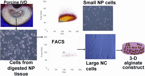
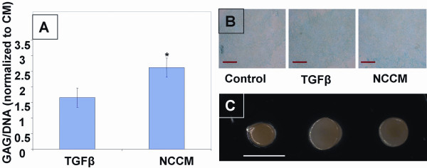
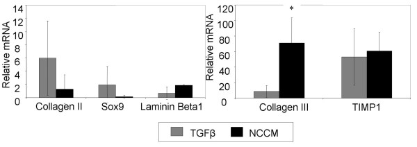
Methods: Human MSCs from bone marrow were expanded and pelleted to form high-density cultures. MSC pellets were exposed to either control medium (CM), chondrogenic medium (CM with dexamethasone and transforming growth factor, (TGF)-beta3) or notochordal cell conditioned medium (NCCM). NCCM was prepared from NC cells maintained in serum free medium for four days. After seven days culture, MSC pellets were analyzed for appearance, biochemical composition (glycosaminoglycans and DNA), and gene expression profile (sox-9, collagen types-II and III, laminin-beta1 and TIMP1(tissue inhibitor of metalloproteinases-1)).
Results: Significantly higher glycosaminoglycan accumulation was seen in NCCM treated pellets than in CM or TGFbeta groups. With NCCM treatment, increased gene expression of collagen III, and a trend of increasing expression of laminin-beta1 and decreased expression of sox-9 and collagen II relative to the TGFbeta group was observed.
Conclusions: Together, results suggest NCCM stimulates mesenchymal stem cell differentiation toward a potentially NP-like phenotype with some characteristics of the developing IVD.
Figures
Similar articles
-
Notochordal conditioned media from tissue increases proteoglycan accumulation and promotes a healthy nucleus pulposus phenotype in human mesenchymal stem cells.Arthritis Res Ther. 2011 May 31;13(3):R81. doi: 10.1186/ar3344. Arthritis Res Ther. 2011. PMID: 21627827 Free PMC article.
-
Notochordal cells protect nucleus pulposus cells from degradation and apoptosis: implications for the mechanisms of intervertebral disc degeneration.Arthritis Res Ther. 2011;13(6):R215. doi: 10.1186/ar3548. Epub 2011 Dec 29. Arthritis Res Ther. 2011. PMID: 22206702 Free PMC article.
-
Conditioned medium derived from notochordal cell-rich nucleus pulposus tissue stimulates matrix production by canine nucleus pulposus cells and bone marrow-derived stromal cells.Tissue Eng Part A. 2015 Mar;21(5-6):1077-84. doi: 10.1089/ten.TEA.2014.0309. Epub 2014 Dec 17. Tissue Eng Part A. 2015. PMID: 25370929 Free PMC article.
-
Growth and differentiation factor-5 contributes to the structural and functional maintenance of the intervertebral disc.Cell Physiol Biochem. 2015;35(1):1-16. doi: 10.1159/000369670. Epub 2015 Jan 2. Cell Physiol Biochem. 2015. PMID: 25547527 Review.
-
Current Status of the Instructional Cues Provided by Notochordal Cells in Novel Disc Repair Strategies.Int J Mol Sci. 2021 Dec 31;23(1):427. doi: 10.3390/ijms23010427. Int J Mol Sci. 2021. PMID: 35008853 Free PMC article. Review.
Cited by
-
The cell-based approach in neurosurgery: ongoing trends and future perspectives.Heliyon. 2019 Nov 26;5(11):e02818. doi: 10.1016/j.heliyon.2019.e02818. eCollection 2019 Nov. Heliyon. 2019. PMID: 31844735 Free PMC article. Review.
-
Photocrosslinkable laminin-functionalized polyethylene glycol hydrogel for intervertebral disc regeneration.Acta Biomater. 2014 Mar;10(3):1102-11. doi: 10.1016/j.actbio.2013.11.013. Epub 2013 Nov 25. Acta Biomater. 2014. PMID: 24287160 Free PMC article.
-
An insight on the N-glycome of notochordal cell-rich porcine nucleus pulposus during maturation.FASEB Bioadv. 2023 Jun 8;5(8):321-335. doi: 10.1096/fba.2023-00011. eCollection 2023 Aug. FASEB Bioadv. 2023. PMID: 37554546 Free PMC article.
-
A Review: Methodologies to Promote the Differentiation of Mesenchymal Stem Cells for the Regeneration of Intervertebral Disc Cells Following Intervertebral Disc Degeneration.Cells. 2023 Aug 28;12(17):2161. doi: 10.3390/cells12172161. Cells. 2023. PMID: 37681893 Free PMC article. Review.
-
Human umbilical cord mesenchymal stromal cells exhibit immature nucleus pulposus cell phenotype in a laminin-rich pseudo-three-dimensional culture system.Stem Cell Res Ther. 2013 Oct 2;4(5):120. doi: 10.1186/scrt331. Stem Cell Res Ther. 2013. PMID: 24405888 Free PMC article.
References
Publication types
MeSH terms
Substances
Grants and funding
LinkOut - more resources
Full Text Sources
Other Literature Sources
Research Materials
Miscellaneous
