. 2010;12(3):R118.
doi: 10.1186/ar3055. Epub 2010 Jun 16.

Interleukin-18 as an in vivo mediator of monocyte recruitment in rodent models of rheumatoid arthritis

Affiliations

Interleukin-18 as an in vivo mediator of monocyte recruitment in rodent models of rheumatoid arthritis

Jeffrey H Ruth et al. Arthritis Res Ther. 2010.

Abstract

Introduction: The function of interleukin-18 (IL-18) was investigated in pertinent animal models of rodent rheumatoid arthritis (RA) to determine its proinflammatory and monocyte recruitment properties.

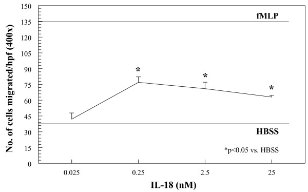
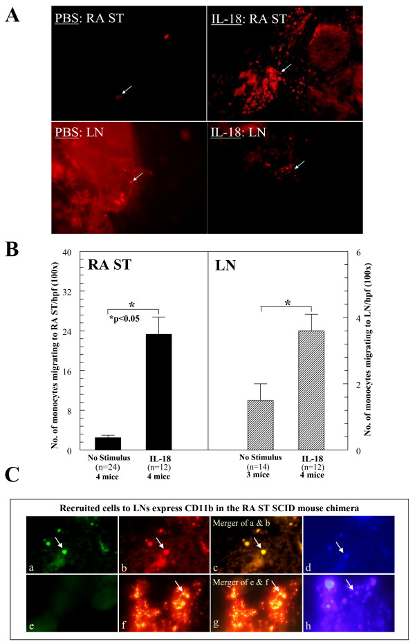
Methods: We used a modified Boyden chemotaxis system to examine monocyte recruitment to recombinant human (rhu) IL-18 in vitro. Monocyte recruitment to rhuIL-18 was then tested in vivo by using an RA synovial tissue (ST) severe combined immunodeficient (SCID) mouse chimera. We defined monocyte-specific signal-transduction pathways induced by rhuIL-18 with Western blotting analysis and linked this to in vitro monocyte chemotactic activity. Finally, the ability of IL-18 to induce a cytokine cascade during acute joint inflammatory responses was examined by inducing wild-type (Wt) and IL-18 gene-knockout mice with zymosan-induced arthritis (ZIA).

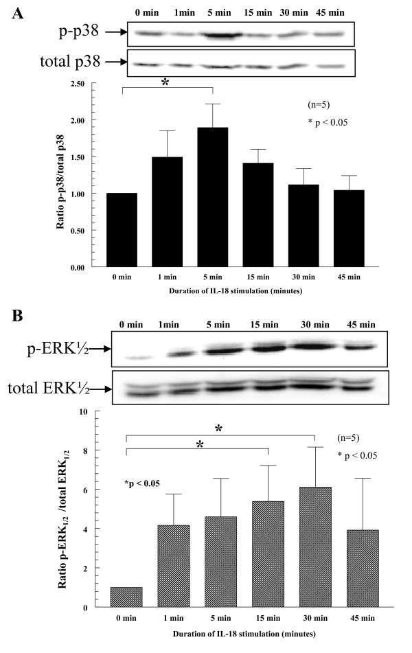
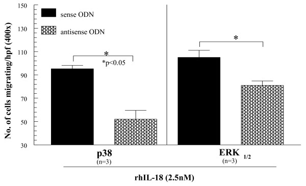
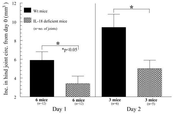
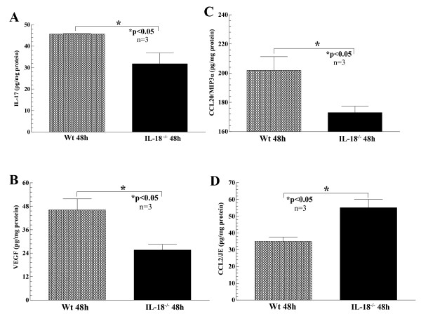
Results: We found that intragraft injected rhuIL-18 was a robust monocyte recruitment factor to both human ST and regional (inguinal) murine lymph node (LN) tissue. IL-18 gene-knockout mice also showed pronounced reductions in joint inflammation during ZIA compared with Wt mice. Many proinflammatory cytokines were reduced in IL-18 gene-knockout mouse joint homogenates during ZIA, including macrophage inflammatory protein-3alpha (MIP-3alpha/CCL20), vascular endothelial cell growth factor (VEGF), and IL-17. Signal-transduction experiments revealed that IL-18 signals through p38 and ERK1/2 in monocytes, and that IL-18-mediated in vitro monocyte chemotaxis can be significantly inhibited by disruption of this pathway.

Conclusions: Our data suggest that IL-18 may be produced in acute inflammatory responses and support the notion that IL-18 may serve a hierarchic position for initiating joint inflammatory responses.


Figures

Figure 1
Figure 1
Monocytes were isolated from the peripheral blood (PB) of normal (NL) volunteers and placed in a modified Boyden chemotaxis system opposite graded increases in concentration of rhuIL-18. As shown, IL-18 stimulates chemotaxis for human monocytes in a dose-dependent manner, and is maximal between 0.25 nM and 25 nM (figure representative of three separate experiments).
Figure 2
Figure 2
IL-18 activates p-p38 and p-ERK½ in a time-dependent manner. Monocytes (5 × 10⁶ cells) were stimulated with 2.5 nM rhuIL-18. Cell lysates were made and probed for p-p38 and p-ERK½ with Western blot, showing marked increases in phosphorylation after 5 minutes for p-p38 and 15 to 30 minutes for p-ERK½. Representative blots show both p-p38 and p-ERK½ (upper panel for p-p38 and lower panel for p-ERK½). Graphs for p-p38 and p-ERK½ were normalized by respective total cellular expression for both signaling molecules relative to the untreated control blots (n = the number of blood donors, and graphs show combined data from five separate experiments). In total, five separate experiments were completed by using peripheral blood monocytes from four separate volunteers.
Figure 3
Figure 3
Monocytes were suspended at 2.5 × 10⁶ cells/ml and then transfected with sense or antisense ODNs in serum-free media for 4 hours. Transfection efficiency for all genes was routinely > 80%, as determined by counting fluorescein isothiocyanate (FITC)-transfected cells with fluorescence microscopy and comparing with a DAPI label in the same cells (data not shown). Transfected cells were added to Boyden chemotaxis chambers to determine their migratory activity toward rhuIL-18 (2.5 nM). As shown, monocytes transfected with either antisense p38 or ERK½ showed significant reductions in chemotaxic activity toward rhuIL-18 compared with sense transfected cells (n = number of experimental repeats from independent PB monocyte donors).
Figure 4
Figure 4
Peripheral blood monocytes injection. (A) PKH26 red fluorescent dye-tagged human peripheral blood (PB) monocytes (5 × 10⁶) were injected i.v. into SCID mice engrafted for 4 to 6 weeks with human rheumatoid arthritis synovial tissue (RA ST). Before administering cells, ST grafts were injected with rhuIL-18 (1,000 ng/graft) or sham injected (PBS stimulus). At 48 hours, grafts and inguinal lymph nodes (LNs) were harvested, and tissue sections were examined with immunofluorescence microscopy at 550 nm (100 ×). The top panel shows PKH26 dye-tagged monocytes migrating into PBS or rhuIL-18 injected RA ST. (B) The lower portion of the same panel shows an image of the local LNs containing recruited monocytes from the same mice. The number of dye-tagged cells migrating to engrafted RA ST or LN tissue in response to rhuIL-18 is graphed in the next panel. As shown, SCID mice receiving intragraft injections of rhuIL-18 showed significant recruitment of human monocytes to both engrafted RA ST and murine LNs. Monocyte migration was quantified by dividing the number of cells per hpf/tissue section at 100 × (n = number of tissue sections counted ± SEM). (C) LNs from rhuIL-18 simulated SCID chimeric mice were harvested and evaluated for human monocyte recruitment. LNs were stained for CD11b/Mac-1 with fluorescence histology. The primary antibody was a mouse anti-human mAb, followed by blocking with goat serum and the addition of a goat anti-mouse FITC-tagged secondary antibody. (a) Human monocytes expressing CD11b/Mac-1 migrate to murine LNs (fluorescent green cells, see arrow). (b) Fluorescent dye-tagged human cells in murine LNs. (c) Merger of (a) and (b), showing that the migrating cells are expressing human CD11b/Mac-1 (fluorescent yellow staining; see arrow). (d) DAPI staining showing cell nuclei (fluorescent blue cells, see arrow). (e) Negative-control staining for CD11b/Mac-1 (nonspecific IgG was used as the primary mAb). (f) Murine LN showing recruited cells (red fluorescent staining, see arrow). (g) Merger of (e) and (f) showing a lack of nonspecific cellular staining. (h) DAPI staining showing cell nuclei (original magnification, 400 ×).
Figure 5
Figure 5
Wt and IL-18 gene-knockout mice were administered zymosan to induce zymosan-induced arthritis (ZIA). Wt mice showed increases of hind joint (knee) circumference from 24 to 48 hours, with a pronounced reduction of swelling in comparative mice lacking IL-18. These data show that IL-18 is critical in acute inflammation of murine joints in as early as 24 hours after zymosan injection (n = number of joints analyzed).
Figure 6
Figure 6
Joint homogenates were prepared from both Wt and IL-18 gene-knockout mice injected with zymosan to induce zymosan-induced arthritis (ZIA). All tissue homogenates were initially measured for total protein content to normalize cytokine expression to total protein content for comparison between cytokines. Cytokines measured included IL-1β IL-6, IL-17, TNF-α MCP-1/CCL2, MIP-1α/CCL3, MIP-3α/CCL20, RANTES/CCL5, and VEGF. Although all cytokines measured were detectable in all the tissue homogenates, significant decreases of IL-17 (a), VEGF (b), and MIP-3α/CCL20 (c) were found in the IL-18 gene-knockout homogenates compared with Wt mice. Conversely, MCP-1/CCL2 (d) was significantly increased in the same homogenates from IL-18 gene-knockout compared with Wt mice (n = number of joints examined)

References

    1. Boraschi D, Dinarello CA. IL-18 in autoimmunity: review. Eur Cytokine Netwk. 2006;17:224–252. - PubMed
    1. Kawashima M, Yamamura M, Taniai M, Yamauchi H, Tanimoto T, Kurimoto M, Miyawaki S, Amano T, Takeuchi T, Makino H. Levels of interleukin-18 and its binding inhibitors in the blood circulation of patients with adult-onset Still's disease. Arthritis Rheum. 2001;44:550–560. doi: 10.1002/1529-0131(200103)44:3<550::AID-ANR103>3.0.CO;2-5. - DOI - PubMed
    1. Moller B, Kukoc-Zivojnov N, Kessler U, Rehart S, Kaltwasser JP, Hoelzer D, Kalina U, Ottmann OG. Expression of interleukin-18 and its monokine-directed function in rheumatoid arthritis. Rheumatology. 2001;40:302–309. doi: 10.1093/rheumatology/40.3.302. - DOI - PubMed
    1. Bresnihan B, Roux-Lombard P, Murphy E, Kane D, FitzGerald O, Dayer JM. Serum interleukin 18 and interleukin 18 binding protein in rheumatoid arthritis. Ann Rheum Dis. 2002;61:726–729. doi: 10.1136/ard.61.8.726. - DOI - PMC - PubMed
    1. Gracie JA, Forsey RJ, Chan WL, Gilmour A, Leung BP, Greer MR, Kennedy K, Carter R, Wei XQ, Xu D, Field M, Foulis A, Liew FY, McInnes IB. A proinflammatory role for IL-18 in rheumatoid arthritis. J Clin Invest. 1999;104:1393–1401. doi: 10.1172/JCI7317. - DOI - PMC - PubMed

Publication types

MeSH terms

Substances