The Src inhibitor dasatinib accelerates the differentiation of human bone marrow-derived mesenchymal stromal cells into osteoblasts
- PMID: 20565769
- PMCID: PMC3087319
- DOI: 10.1186/1471-2407-10-298
The Src inhibitor dasatinib accelerates the differentiation of human bone marrow-derived mesenchymal stromal cells into osteoblasts
Expression of concern in
-
Editorial Expression of Concern: The Src inhibitor dasatinib accelerates the differentiation of human bone marrow-derived mesenchymal stromal cells into osteoblasts.BMC Cancer. 2025 Jan 9;25(1):49. doi: 10.1186/s12885-025-13460-1. BMC Cancer. 2025. PMID: 39789513 Free PMC article. No abstract available.
Abstract
Background: The proto-oncogene Src is an important non-receptor protein tyrosine kinase involved in signaling pathways that control cell adhesion, growth, migration and differentiation. It negatively regulates osteoblast activity, and, as such, its inhibition is a potential means to prevent bone loss. Dasatinib is a new dual Src/Bcr-Abl tyrosine kinase inhibitor initially developed for the treatment of chronic myeloid leukemia. It has also shown promising results in preclinical studies in various solid tumors. However, its effects on the differentiation of human osteoblasts have never been examined.
Methods: We evaluated the effects of dasatinib on bone marrow-derived mesenchymal stromal cells (MSC) differentiation into osteoblasts, in the presence or absence of a mixture of dexamethasone, ascorbic acid and beta-glycerophosphate (DAG) for up to 21 days. The differentiation kinetics was assessed by evaluating mineralization of the extracellular matrix, alkaline phosphatase (ALP) activity, and expression of osteoblastic markers (receptor activator of nuclear factor kappa B ligand [RANKL], bone sialoprotein [BSP], osteopontin [OPN]).
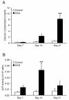
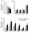
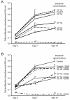
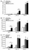
Results: Dasatinib significantly increased the activity of ALP and the level of calcium deposition in MSC cultured with DAG after, respectively, 7 and 14 days; it upregulated the expression of BSP and OPN genes independently of DAG; and it markedly downregulated the expression of RANKL gene and protein (decrease in RANKL/OPG ratio), the key factor that stimulates osteoclast differentiation and activity.
Conclusions: Our results suggest a dual role for dasatinib in both (i) stimulating osteoblast differentiation leading to a direct increase in bone formation, and (ii) downregulating RANKL synthesis by osteoblasts leading to an indirect inhibition of osteoclastogenesis. Thus, dasatinib is a potentially interesting candidate drug for the treatment of osteolysis through its dual effect on bone metabolism.
Figures



Similar articles
-
Src family kinase/abl inhibitor dasatinib suppresses proliferation and enhances differentiation of osteoblasts.Oncogene. 2010 Jun 3;29(22):3196-207. doi: 10.1038/onc.2010.73. Epub 2010 Mar 15. Oncogene. 2010. PMID: 20228840 Free PMC article.
-
A novel collagen-binding peptide promotes osteogenic differentiation via Ca2+/calmodulin-dependent protein kinase II/ERK/AP-1 signaling pathway in human bone marrow-derived mesenchymal stem cells.Cell Signal. 2008 Apr;20(4):613-24. doi: 10.1016/j.cellsig.2007.11.012. Epub 2007 Nov 29. Cell Signal. 2008. PMID: 18248957
-
Dasatinib as a bone-modifying agent: anabolic and anti-resorptive effects.PLoS One. 2012;7(4):e34914. doi: 10.1371/journal.pone.0034914. Epub 2012 Apr 23. PLoS One. 2012. PMID: 22539950 Free PMC article.
-
Role of farnesoid X receptor (FXR) in the process of differentiation of bone marrow stromal cells into osteoblasts.Bone. 2011 Dec;49(6):1219-31. doi: 10.1016/j.bone.2011.08.013. Epub 2011 Aug 26. Bone. 2011. PMID: 21893226
-
Targeting SRC in glioblastoma tumors and brain metastases: rationale and preclinical studies.Cancer Lett. 2010 Dec 8;298(2):139-49. doi: 10.1016/j.canlet.2010.08.014. Cancer Lett. 2010. PMID: 20947248 Free PMC article. Review.
Cited by
-
Mechanical loading in osteocytes induces formation of a Src/Pyk2/MBD2 complex that suppresses anabolic gene expression.PLoS One. 2014 May 19;9(5):e97942. doi: 10.1371/journal.pone.0097942. eCollection 2014. PLoS One. 2014. PMID: 24841674 Free PMC article.
-
Dasatinib accelerates valproic acid-induced acute myeloid leukemia cell death by regulation of differentiation capacity.PLoS One. 2014 Jun 11;9(2):e98859. doi: 10.1371/journal.pone.0098859. eCollection 2014. PLoS One. 2014. PMID: 24918603 Free PMC article.
-
1,25-Dihydroxyvitamin D3 prevents bone loss of the secondary spongiosa in arthritic rats by an increase of bone formation and mineralization and inhibition of bone resorption.BMC Musculoskelet Disord. 2014 Oct 14;15:345. doi: 10.1186/1471-2474-15-345. BMC Musculoskelet Disord. 2014. PMID: 25315028 Free PMC article.
-
Role of the GRP78-c-Src signaling pathway on osteoblast differentiation of periodontal ligament fibroblasts induced by cyclic mechanical stretch.Hua Xi Kou Qiang Yi Xue Za Zhi. 2024 Jun 1;42(3):304-312. doi: 10.7518/hxkq.2024.2023354. Hua Xi Kou Qiang Yi Xue Za Zhi. 2024. PMID: 39049649 Free PMC article. Chinese, English.
-
The tyrosine kinase inhibitor dasatinib induces a marked adipogenic differentiation of human multipotent mesenchymal stromal cells.PLoS One. 2011;6(12):e28555. doi: 10.1371/journal.pone.0028555. Epub 2011 Dec 2. PLoS One. 2011. PMID: 22164306 Free PMC article.
References
Publication types
MeSH terms
Substances
LinkOut - more resources
Full Text Sources
Molecular Biology Databases
Research Materials
Miscellaneous

