

Different significance between intratumoral and peritumoral lymphatic vessel density in gastric cancer: a retrospective study of 123 cases
- PMID: 20565772
- PMCID: PMC2906480
- DOI: 10.1186/1471-2407-10-299
Different significance between intratumoral and peritumoral lymphatic vessel density in gastric cancer: a retrospective study of 123 cases
Abstract
Background: Patients with gastric cancer in China have worse outcome and poorer prognosis. Tumor-induced lymphangiogenesis plays a crucial role in metastasis and tumor progression. The intratumoral and peritumoral lymphatics were supposed to have different biological effects. Three major growth factors, vascular endothelial growth factor- (VEGF)-A, VEGF-C and VEGF-D, are involved in the activation process via their receptors (VEGFRs). The purpose of current study is to investigate the significant difference between intratumoral and peritumoral lymphatic vessel density (LVD) in gastric cancer and their correlations with lymphangiogenetic growth factors.
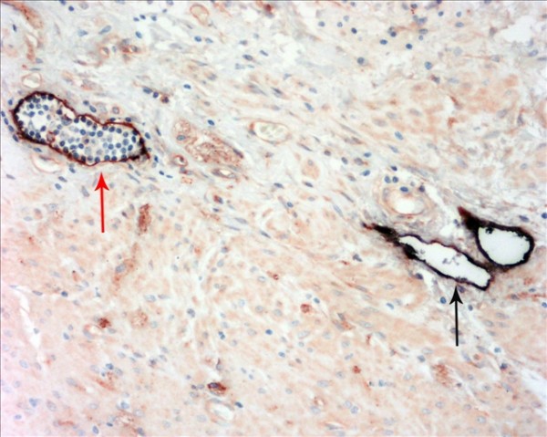
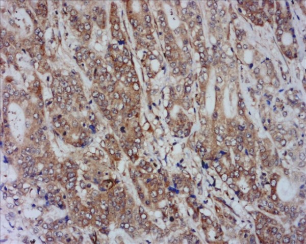
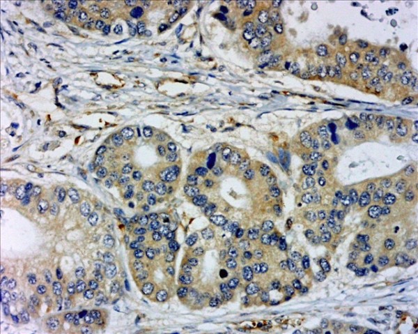
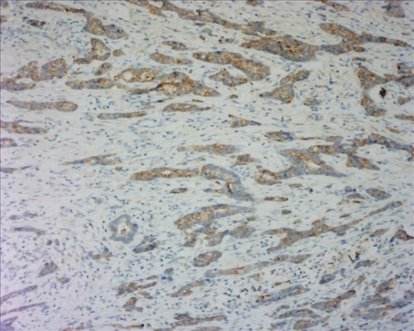
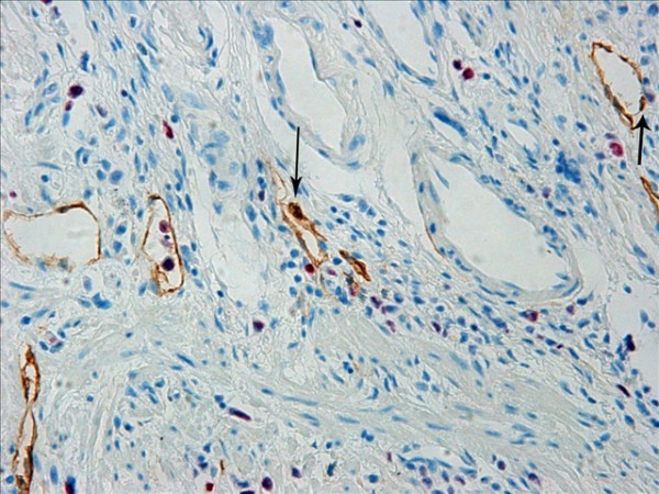
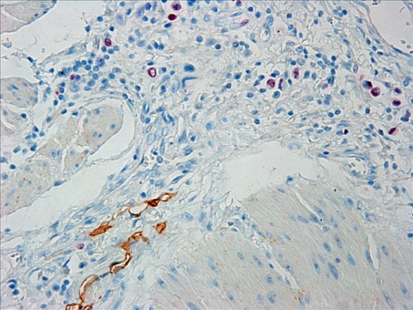
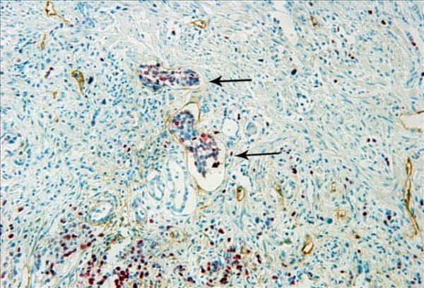
Methods: Intratumoral LVD (I-LVD) and peritumoral LVD (P-LVD) of 123 patients with primary gastric cancer were assessed after staining with D2-40, and confirmed by double staining with D2-40/CD34. Proliferative activity of lymphatics endothelium was evaluated by double staining with D2-40/Ki-67. The associations were analyzed between I-LVD/P-LVD and the expression level of VEGF-A, VEGF-C, VEGF-D and the receptor VEGFR-3, which was measured by immunohistochemistry (IHC). The correlations of I-LVD and P-LVD with patient prognosis were also valued.
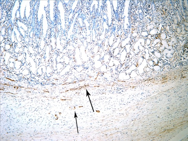
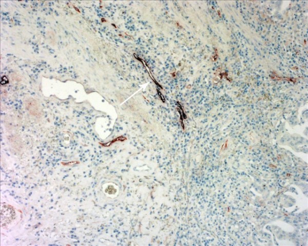
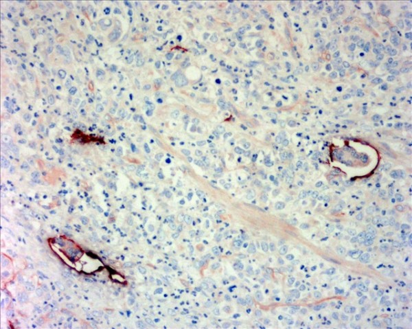
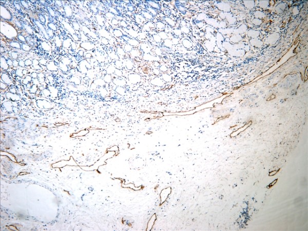
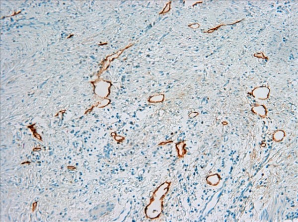
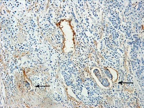
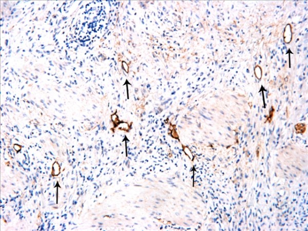
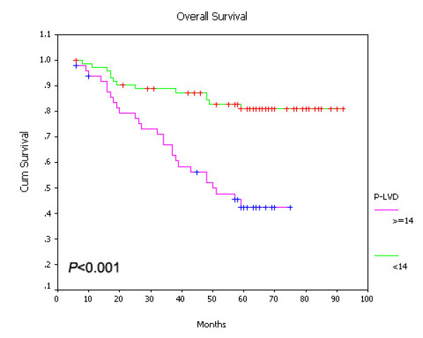
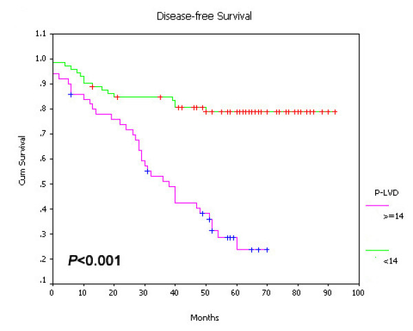
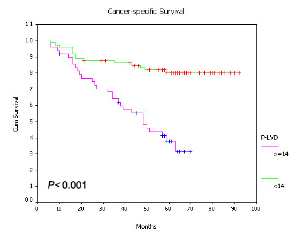
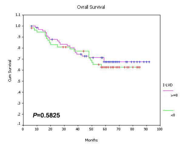
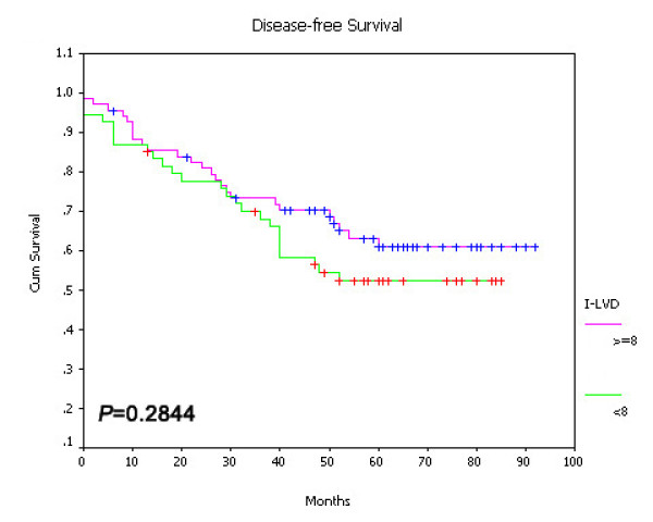
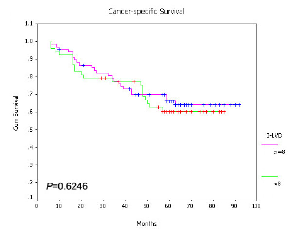
Results: (1) The peritumoral lymphatics (PTLs) were relatively enlarged with dilated lumen compared with the intratumoral lymphatics (ITLs). Increased P-LVD was significantly higher than I-LVD (P < 0.05). (2) P-LVD was found significantly associated with lymph node metastasis (LNM) (P < 0.001), lymphatic vessel invasion (LVI) (P < 0.001), VEGF-C (P = 0.003), VEGF-D expression level (P = 0.005) and VEGFR-3 expression level (P < 0.001) in peritumoral tissues, despite no significant association was found between above variants with I-LVD. However, increased I-LVD was demonstrated to be associated with decreased tumor volume (P < 0.001). Neither I-LVD nor P-LVD was correlated with VEGF-A expression (P > 0.05). (3) Proliferative activity of lymphatics endothelium was observed in PTLs, in spite of ITLs. (4) Increased P-LVD, but not I-LVD, was indicated to be an independent risk factor for lymph node metastasis by multivariate logistic regression analysis, and was related to worse disease-free survival and overall survival.
Conclusions: PTLs play roles in gastric cancer progression. Increased P-LVD, but not I-LVD, was significantly associated with VEGF-C/-D/VEGFR-3 system, and could be an independent risk factor for lymph node metastasis and a prognostic factor in gastric cancer.
Figures
Similar articles
-
Peritumoral lymphangiogenesis induced by vascular endothelial growth factor C and D promotes lymph node metastasis in breast cancer patients.World J Surg Oncol. 2012 Aug 20;10:165. doi: 10.1186/1477-7819-10-165. World J Surg Oncol. 2012. PMID: 22906075 Free PMC article.
-
Expressions of COX-2 and VEGF-C in gastric cancer: correlations with lymphangiogenesis and prognostic implications.J Exp Clin Cancer Res. 2011 Jan 28;30(1):14. doi: 10.1186/1756-9966-30-14. J Exp Clin Cancer Res. 2011. PMID: 21272377 Free PMC article.
-
Lymphangiogenesis in gastric carcinoma correlates with prognosis.J Pathol. 2009 Jun;218(2):192-200. doi: 10.1002/path.2523. J Pathol. 2009. PMID: 19224540
-
Lymphangiogenesis and colorectal cancer.Saudi Med J. 2017 Mar;38(3):237-244. doi: 10.15537/smj.2017.3.16245. Saudi Med J. 2017. PMID: 28251217 Free PMC article. Review.
-
Lymphangiogenesis in Breast Cancer: From Molecular Mechanisms to Clinical Implications.FASEB J. 2025 May 15;39(9):e70590. doi: 10.1096/fj.202500024R. FASEB J. 2025. PMID: 40320983 Review.
Cited by
-
Prognostic significance of vascular endothelial growth factor immunohistochemical expression in gastric cancer: a meta-analysis.Mol Biol Rep. 2012 Oct;39(10):9473-84. doi: 10.1007/s11033-012-1812-8. Epub 2012 Jun 23. Mol Biol Rep. 2012. PMID: 22729879
-
Does immunohistochemical staining have a clinical impact in early gastric cancer conducted endoscopic submucosal dissection?World J Gastroenterol. 2012 Sep 7;18(33):4578-84. doi: 10.3748/wjg.v18.i33.4578. World J Gastroenterol. 2012. PMID: 22969232 Free PMC article.
-
Lymphangiogenesis in gastric cancer regulated through Akt/mTOR-VEGF-C/VEGF-D axis.BMC Cancer. 2015 Mar 7;15:103. doi: 10.1186/s12885-015-1109-0. BMC Cancer. 2015. PMID: 25884175 Free PMC article.
-
Lymphangiogenesis in gastric cancer: function and mechanism.Eur J Med Res. 2023 Oct 7;28(1):405. doi: 10.1186/s40001-023-01298-x. Eur J Med Res. 2023. PMID: 37803421 Free PMC article. Review.
-
VEGF-C and Lymphatic Vessel Density in Tumor Tissue of Gastric Cancer: Correlations with Pathoclinical Features and Prognosis.Cancers (Basel). 2025 Apr 23;17(9):1406. doi: 10.3390/cancers17091406. Cancers (Basel). 2025. PMID: 40361332 Free PMC article.
References
-
- Onogawa S, Kitadai Y, Amioka T, Kodama M, Cho S, Kuroda T, Ochiumi T, Kimura S, Kuwai T, Tanaka S, Chayama K. Expression of vascular endothelial growth factor (VEGF)-C and VEGF-D in early gastric carcinoma: correlation with clinicopathological parameters. Cancer Lett. 2005;226:85–90. doi: 10.1016/j.canlet.2004.12.030. - DOI - PubMed
-
- Hachisuka T, Narikiyo M, Yamada Y, Ishikawa H, Ueno M, Uchida H, Yoriki R, Ohigashi Y, Miki K, Tamaki H, Mizuno T, Nakajima Y. High lymphatic vessel density correlates with overexpression of VEGF-C in gastric cancer. Oncol Rep. 2005;13:733–737. - PubMed
Publication types
MeSH terms
Substances
LinkOut - more resources
Full Text Sources
Medical
Miscellaneous
