. 2010 Jun 18:5:15.
doi: 10.1186/1749-8104-5-15.

Assembly of the outer retina in the absence of GABA synthesis in horizontal cells

Affiliations

Assembly of the outer retina in the absence of GABA synthesis in horizontal cells

Timm Schubert et al. Neural Dev. .

Abstract

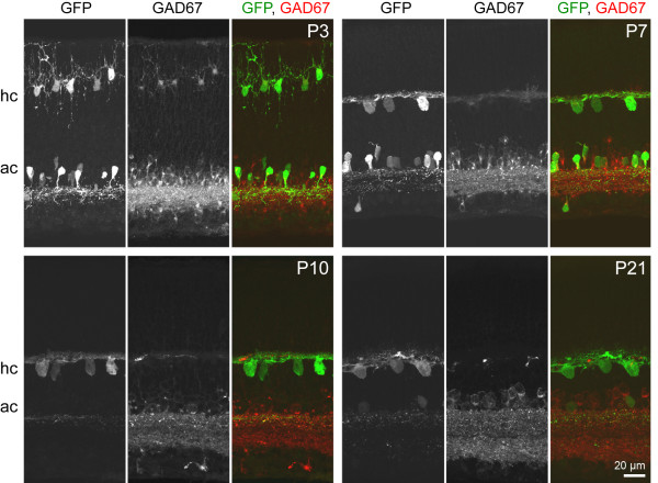
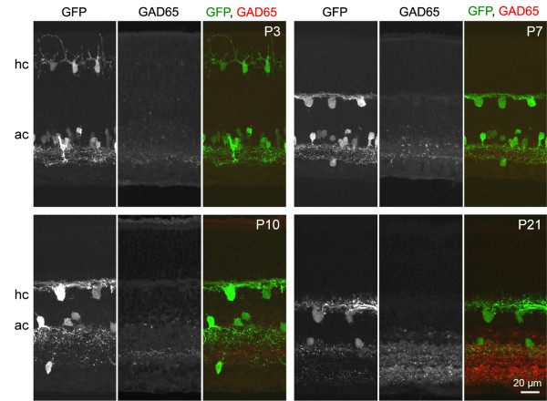
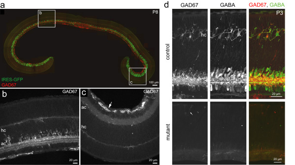
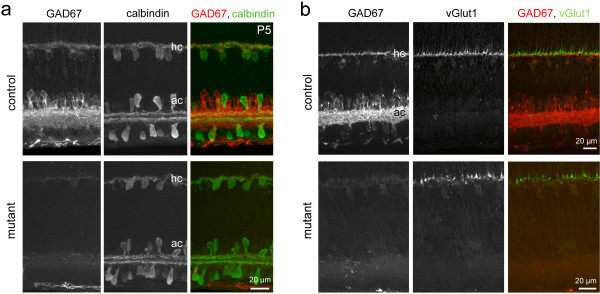
Background: The inhibitory neurotransmitter gamma-amino-butyric acid (GABA) not only modulates excitability in the mature nervous system but also regulates neuronal differentiation and circuit development. Horizontal cells, a subset of interneurons in the outer retina, are transiently GABAergic during the period of cone photoreceptor synaptogenesis. In rodents, both horizontal cells and cone axonal terminals express GABAA receptors. To explore the possibility that transient GABA expression in mouse neonatal horizontal cells influences the structural development of synaptic connectivity in the outer retina, we examined a mutant in which expression of GAD67, the major synthesizing enzyme for GABA, is selectively knocked out in the retina.

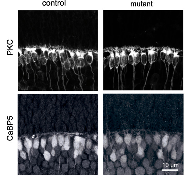
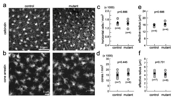
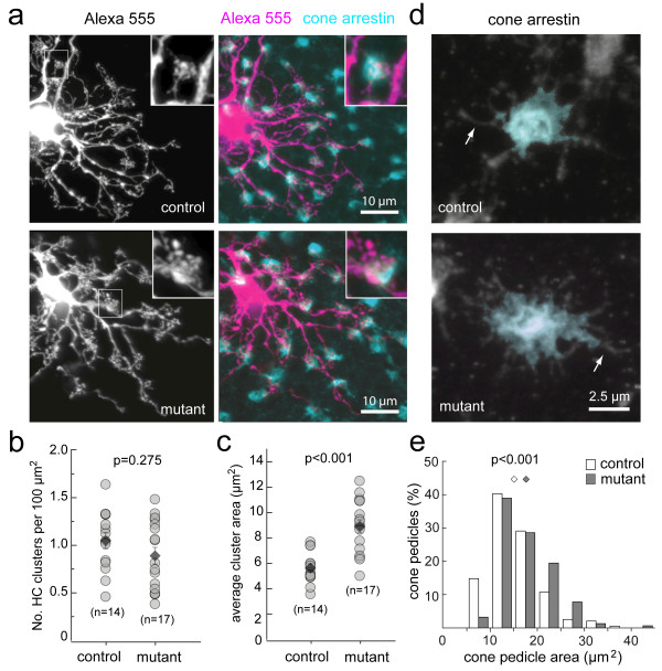
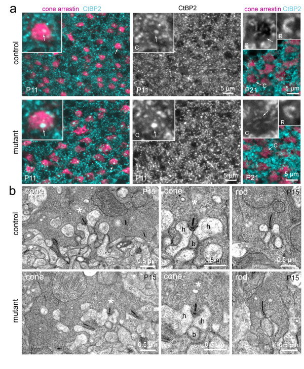
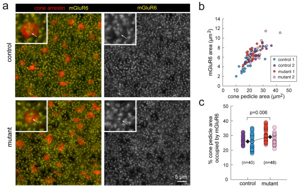
Results: Immunocytochemistry and electron microscopy revealed that the assembly of triad synapses involving cone axonal pedicles and the dendrites of horizontal and bipolar cells is unaffected in the mutant retina. Moreover, loss of GABA synthesis in the outer retina did not perturb the spatial distributions and cell densities of cones and horizontal cells. However, there were some structural alterations at the cellular level: the average size of horizontal cell dendritic clusters was larger in the mutant, and there was also a small but significant increase in cone photoreceptor pedicle area. Moreover, metabotropic glutamate receptor 6 (mGluR6) receptors on the dendrites of ON bipolar cells occupied a slightly larger proportion of the cone pedicle in the mutant.

Conclusions: Together, our analysis shows that transient GABA synthesis in horizontal cells is not critical for synapse assembly and axonal and dendritic lamination in the outer retina. However, pre- and postsynaptic structures are somewhat enlarged in the absence of GABA in the developing outer retina, providing for a modest increase in potential contact area between cone photoreceptors and their targets. These findings differ from previous results in which pharmacological blockade of GABAA receptors in the neonatal rabbit retina caused a reduction in cone numbers and led to a grossly disorganized outer retina.


Figures

Figure 1
Figure 1
Horizontal cells transiently express GAD67 during early postnatal development. Retinal cross-sections from the G42 mouse in which horizontal cells (hc) and amacrine cells (ac) expressing GFP were immunolabeled for GAD67 during early postnatal development (P3 to P10) and at structural maturity (P21).
Figure 2
Figure 2
Horizontal cells do not express GAD65 at any stage of postnatal development. Retinal cross-sections from neonatal G42 mice immunolabeled for GAD65. hc, horizontal cells; ac, amacrine cells.
Figure 3
Figure 3
Pattern of GAD67 expression in αPax6-Cre:Gad1lox/lox (mutant) retina. (a) The spatial distribution of Cre recombinase expression across the retina parallels the pattern of GFP expression in amacrine cells. Cre expression is present in all cells of the embryonic retina, but several days after birth becomes restricted to subpopulations of amacrine cells [63]. At P8, GFP is expressed by relatively few cells in central retina (box, b) and a larger number of amacrine cells in the peripheral retina (box, c). Staining for GAD67 follows an inverse pattern in this mutant retina. (b) Numerous GAD67-immunopositive amacrine cells near the center of the mutant retina. hc, horizontal cells; ac, amacrine cells. (c) GAD67 immunoreactivity is largely absent in the retinal periphery of the mutant retina. Immunofluorescence in this image (arrow) marks blood vessels. There is no GAD67 immunoreactivity at retinal depths where amacrine cells (ac) and horizontal cells (hc) are located. (d) In the wild-type region of a P3 mutant retina, GAD67-immunopositive horizontal cells (hc) also contain GABA. In knockout regions of this retina, GAD67 and GABA immunolabeling are absent. Arrow (mutant) indicates immunoreactivity in some blood vessels.
Figure 4
Figure 4
Horizontal cell and photoreceptor lamination are not grossly disrupted in the absence of GABA in the outer retina. (a) Horizontal cells from a P5 mutant retina immunolabeled for calbindin. In both littermate control and mutant retinas, the processes of these cells are stratified within the outer retina. (b) Photoreceptor terminals in a P5 mutant retina immunostained for vesicular glutamate transporter 1 (vGluT1). The terminals are stratified in both littermate control and mutant retinas. The slight irregularity of the mutant vGlut1 lamination is not unusual at this age and can be observed in the wild-type retina as well. hc, horizontal cells; ac, amacrine cells.
Figure 5
Figure 5
Bipolar cell dendrites are stratified in the absence of GABA synthesis by horizontal cells. P10 rod bipolar cells immunolabeled for PKC and cone bipolar cells immunolabeled for CaBP5 have stratified dendritic arbors in the mutant retina.
Figure 6
Figure 6
Density and distribution of horizontal cells and cones are normal in the mutant retina. (a,b) Populations of horizontal cells (a) and cone pedicles (b) immunostained for calbindin and cone-arrestin, respectively, for P11 retinas. (c,d) Plots of the density of horizontal cells and cones for littermate controls and mutant retinas. (e,f) Comparison of effective radii for horizontal cells and cones in littermate controls and mutant retinas. (c-f) n = number of retinas; filled diamond and error bar = mean and standard error.
Figure 7
Figure 7
Horizontal cell dendritic cluster area and cone pedicle size are significantly increased in the mutant retina. (a) Examples of horizontal cells in P10 littermate control and mutant retinas that were intracellularly dye-filled with Alexa Fluor 555. Mutant retinas were immunostained for GAD67 to determine whether cells were located in the wild-type or knockout regions (not shown). Insets show dendritic terminal clusters that overlay cone pedicles (revealed by immunostaining for cone-arrestin) at their sites of contact. (b,c) Quantification of horizontal cell (HC) terminal cluster density (number of clusters per 100 μm2 dendritic field area) and average cluster area per cell. n = number of retinas. Filled diamond and error bar = mean and standard error. (d) High magnification examples of the maximum intensity projections of an image stack through a cone pedicle in a littermate control retina and a pedicle in the mutant retina. Colored regions within the cone pedicles mark the cone pedicle 'area' determined from their respective labeled-fields (see Materials and methods). Arrows indicate an example of filopodia that decorate the base of the cone pedicles. (e) Distributions of cone pedicle areas in littermate control and mutant retinas. Diamonds indicate means of the distributions: mutant (17.15 ± 0.46 μm2), n = 154 pedicles, n = 4 animals; control (15.20 ± 0.38 μm2), n = 196 pedicles, 5 animals.
Figure 8
Figure 8
Ribbons are present and localized to synaptic sites in the mutant retina. (a) Immunostaining for cone pedicles (cone-arrestin) and ribbons (C-terminus binding protein 2 (CtBP2)) in littermate controls and knockout regions of the mutant retina. Insets show higher magnification of cone pedicles marked by arrows in merged images. Arrows in insets indicate bright CtBP2 immunoreactivity - note more rounded structures at P11 compared to ribbon-like structures at P21 for cones. C, cone ribbons. Additionally, higher magnifications of CtBP2 immunostained horseshoe-like ribbons in rod terminals (R) are shown for the P21 retinas. (b) Examples of the ultrastructure of a cone and rod pedicles in control and mutant retinas showing localization of ribbons at synaptic triads. For each of the cone examples, the asterisk indicates the same triad shown in the left and middle columns (h, horizontal cell process; b, bipolar cell dendrite).
Figure 9
Figure 9
Total postsynaptic metabotropic glutamate receptor area within cone pedicles is increased in the mutant retina. (a) Co-labeling of cone pedicles (cone-arrestin) and metabotropic glutamate receptor 6 (mGluR6) on ON bipolar cell dendritic tips at P11. Insets show higher magnification images of the cone pedicles and mGluR6 receptor clusters (arrows). mGluR6 immunoreactivity outside the cone pedicles is associated with rod photoreceptor terminals. (b) Within each cone pedicle, a threshold was applied to the mGluR6 signal and the total area of pixels with intensities above the threshold is plotted against pedicle area. (c) For each cone, the total pixel area corresponding to mGluR6 signal above threshold is presented as a percentage of the area of that cone pedicle. n = number of pedicles analyzed, two animals each for control (three fields) and mutant retinas (three fields).

Similar articles

Cited by

References

    1. Owens DF, Kriegstein AR. Is there more to GABA than synaptic inhibition? Nat Rev Neurosci. 2002;3:715–727. doi: 10.1038/nrn919. - DOI - PubMed
    1. Represa A, Ben-Ari Y. Trophic actions of GABA on neuronal development. Trends Neurosci. 2005;28:278–283. doi: 10.1016/j.tins.2005.03.010. - DOI - PubMed
    1. Bortone D, Polleux F. KCC2 expression promotes the termination of cortical interneuron migration in a voltage-sensitive calcium-dependent manner. Neuron. 2009;62:53–71. doi: 10.1016/j.neuron.2009.01.034. - DOI - PMC - PubMed
    1. Shen W, Da Silva JS, He H, Cline HT. Type A GABA-receptor-dependent synaptic transmission sculpts dendritic arbor structure in Xenopus tadpoles in vivo. J Neurosci. 2009;29:5032–5043. doi: 10.1523/JNEUROSCI.5331-08.2009. - DOI - PMC - PubMed
    1. Chattopadhyaya B, Di Cristo G, Wu CZ, Knott G, Kuhlman S, Fu Y, Palmiter RD, Huang ZJ. GAD67-mediated GABA synthesis and signaling regulate inhibitory synaptic innervation in the visual cortex. Neuron. 2007;54:889–903. doi: 10.1016/j.neuron.2007.05.015. - DOI - PMC - PubMed

Publication types

MeSH terms

Substances

LinkOut - more resources