. 2010 Jun 21:9:156.
doi: 10.1186/1476-4598-9-156.

Inositol 1,4,5-trisphosphate-induced Ca2+ signalling is involved in estradiol-induced breast cancer epithelial cell growth

Affiliations

Inositol 1,4,5-trisphosphate-induced Ca2+ signalling is involved in estradiol-induced breast cancer epithelial cell growth

Cécilia Szatkowski et al. Mol Cancer. .

Abstract

Background: Ca2+ is a ubiquitous messenger that has been shown to be responsible for controlling numerous cellular processes including cell growth and cell death. Whereas the involvement of IP3-induced Ca2+ signalling (IICS) in the physiological activity of numerous cell types is well documented, the role of IICS in cancer cells is still largely unknown. Our purpose was to characterize the role of IICS in the control of growth of the estrogen-dependent human breast cancer epithelial cell line MCF-7 and its potential regulation by 17beta-estradiol (E2).

Results: Our results show that the IP3 receptor (IP3R) inhibitors caffeine, 2-APB and xestospongin C (XeC) inhibited the growth of MCF-7 stimulated by 5% foetal calf serum or 10 nM E2. Furthermore, Ca2+ imaging experiments showed that serum and E2 were able to trigger, in a Ca2+-free medium, an elevation of internal Ca2+ in a 2-APB and XeC-sensitive manner. Moreover, the phospholipase C (PLC) inhibitor U-73122 was able to prevent intracellular Ca2+ elevation in response to serum, whereas the inactive analogue U-73343 was ineffective. Western-blotting experiments revealed that the 3 types of IP3Rs are expressed in MCF-7 cells and that a 48 hours treatment with 10 nM E2 elevated IP3R3 protein expression level in an ICI-182,780 (a specific estrogen receptor antagonist)-dependent manner. Furthermore, IP3R3 silencing by the use of specific small interfering RNA was responsible for a drastic modification of the temporal feature of IICS, independently of a modification of the sensitivity of the Ca2+ release process and acted to counteract the proliferative effect of 10 nM E2.

Conclusions: Altogether, our results are in favour of a role of IICS in MCF-7 cell growth, and we hypothesize that the regulation of IP3R3 expression by E2 is involved in this effect.


Figures
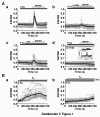
Figure 1
Figure 1
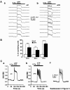
Serum triggers an intracellular Ca2+ signal. (A) The perfusion of a Ca2+-free recording solution containing 5% of serum on Fura-2-loaded MCF-7 cells elicited a strong intracellular Ca2+ signal (a). This signal was due to release from IP3-sensitive Ca2+ stores as it was sensitive to previous application of 2-APB (75 μM, b) or XeC (25 μM, c). Furthermore, U-73122 (20 μM) prevented the effect of serum on intracellular Ca2+ release (d) whereas the inactive analogue U-73343 (20 μM) was ineffective (d, inset). (B) E2 (10 nM) triggered intracellular Ca2+ elevations in MCF-7 cells perfused with a Ca2+-free medium (a); these Ca2+ elevations were inhibited by XeC (25 μM, b). In each panel, the results show the typical traces of 27 to 35 cells, always represented at the same scale; each time, also the mean signal is represented (thick black line).
Figure 2
Figure 2
Pharmacological inhibitors of IP3Rs inhibited 5-FCS- and E2-induced MCF-7 cell growth. (A) The growth of MCF-7 cells induced by a 48 h treatment with 5-FCS was sensitive to pharmacological inhibitors of IP3R. Left bar graph shows the cell number obtained in 0-FCS and serves as a control experiment in order to estimate the proliferative effect of 5-FCS. Caffeine (500 μM) and 2-APB (75 μM) were both able to inhibit significantly 5-FCS-stimulated cell growth (P < 0.05 and P < 0.001, respectively). Values are the mean ± S.E.M. of 6 independent experiments. (B) Pharmacological inhibition of IP3R was responsible for the inhibition of E2-induced cell growth. Whereas caffeine (500 μM) was ineffective alone in control conditions, it inhibited the stimulation of cell growth by E2 (10 nM, P < 0.01). Values are the mean ± S.E.M. of 3 independent experiments. (C) XeC (10 μM) inhibited both 5-FCS and E2-induced MCF-7 cell growth. Values are the mean ± S.E.M. of 4 independent experiments. (D) Kinetics and reversibility of the inhibition by caffeine of E2-induced MCF-7 cell viability. Cells were starved for a 24 h period and were then stimulated by 10 nM E2. Caffeine (500 μM) was either added (+caf) at the beginning of the experiment (a), or after 36 h (b). In both cases, caffeine inhibited the E2-induced increase in cell viability (P < 0.001). The effect of caffeine was reversible since washout (-caf) of this compound after 36 h permitted to significantly restore the proliferative effect of E2 at 72 h (c). Arrows indicate the time of application (downward) or washout (upward) of caffeine. Values are the mean ± S.E.M. of 3 independent experiments.
Figure 3
Figure 3
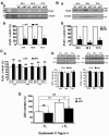
Expression of IP3Rs isoforms and their regulation by E2. (A) MCF-7 cells express the three IP3R isoforms. (a) Western blotting allowed the specific detection in MCF-7 microsomes (50 μg proteins/lane) of IP3R1, IP3R2 and IP3R3. Representative blots for 7 to 8 independent experiments are shown. Whereas E2 had no effect on the expression level of IP3R1 (left panel) and IP3R2 (middle panel), E2 increased the expression of IP3R3 (right panel, P < 0.05). This effect of E2 involves an estrogen receptor-dependent intracellular pathway since it was antagonized by the specific anti-estrogen ICI-182,780 (1 μM, b). (B) Statistical analysis of the effect of E2 on the expression level of the 3 types of IP3Rs (a) and of the inhibitory effect of ICI-182,780 on E2-induced stimulation of IP3R3 expression (b). In the presence or absence of ICI-182,780, the expression level of IP3R3 is not significantly different (113.4 ± 21.9 vs. 100.0 ± 18.2% in control conditions, n = 6).
Figure 4
Figure 4
Silencing of IP3R3 by RNA interference limits the proliferative effect of E2. (A) Validation of the efficiency of the siRNAs used at the mRNA level (a, representative illustrations for 3 independent experiments). The IP3R3 mRNA level was lowered by about 70% (see text for details) at 24, 48 and 72 h post-transfection. (B) siR3 was responsible for a rapid and long-lasting diminution of the expression of IP3R3 at the protein level (a). Compared to control conditions, siR3 diminished the expression of IP3R3 at 24, 48 and 72 h (b). (C) Following IP3R3 silencing, the levels of IP3R1 and IP3R2 mRNA (a) and protein (b) were not significantly changed at 24, 48 and 72 h post-transfection. Representative blots for 3 to 4 independent western blotting experiments are shown. (D) MCF-7 cells were transfected with siC or siR3 and seeded for 18 h in 5-FCS and then starved for 6 h in 0-FCS. Cells were subsequently cultured in 0-FCS in the absence (-E2) of the presence (+E2) of E2 (10 nM) for 48 h. MCF-7 cell number was measured at 72 h post-transfection using the trypan-blue exclusion method. E2-induced increase in cell number was strongly diminished in siR3-transfected MCF-7 cells compared to siC-transfected cells (P < 0.01).
Figure 5
Figure 5
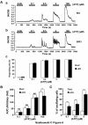
IP3R3 silencing changed the characteristics of ATP-induced Ca2+ signalling in MCF-7 cells. (A) Typical ATP-induced Ca2+ signals in MCF-7 cells 72 h after their transfection with siC (a) or siR3 (b). ATP (5 μM) was perfused in a Ca2+-free recording solution in order to avoid Ca2+ entry. The Ca2+ response changed from a plateau-type pattern to a characteristic oscillatory pattern in siR3-transfected cells. Representative for 6 independent experiments. (B) Statistical analysis of the effect of IP3R3 silencing at 24, 48 and 72 h post-transfection on the percentage of oscillating cells in response to 5 μM ATP in a Ca2+-free recording solution. IP3R3 gene silencing resulted, at any time, in a strong augmentation of the percentage of cells that respond to ATP by an oscillatory Ca2+ signal. (C) Typical ATP-induced Ca2+ signals recorded in a Ca2+-free medium 72 h after transfection with siC (a) or siR3 (b). Ca2+ signals presented in a (full line) and b (dotted line) were superimposed in order to clearly show the modification of the pattern of the Ca2+ response (c).
Figure 6
Figure 6
IP3R3 silencing does not modify the sensitivity of the Ca2+ release process. (A) Typical Ca2+ signals induced by ATP (50 nM - 100 μM) in Fura-2-loaded MCF-7 cells 72 h after their transfection with siC (a) or siR3 (b). ATP triggered intracellular Ca2+ signals with a concentration threshold of about 100 nM in both cell batches but IP3R3 silencing was responsible for a change in the shape of the signal. The percentage of responding cells in siC- and siR3-transfected cells is unchanged whatever the concentration of ATP used (c). Results are representative of 8 independent experiments. (B) The magnitude of the signal is compared at various ATP concentrations (100 nM - 100 μM) and at the maximal concentration of ATP used, the magnitude of the signal is significantly decreased by about 20% (P < 0.01). (C) The number of oscillating cells was investigated at ATP concentrations ranging from 100 nM to 5 μM, and at each concentration the percentage of oscillating cells was largely greater in siR3-transfected cells (P < 0.01 or 0.001).

Similar articles

Cited by

References

    1. Berridge MJ. Calcium signalling and cell proliferation. Bioessays. 1995;17:491–500. doi: 10.1002/bies.950170605. - DOI - PubMed
    1. Berridge MJ, Lipp P, Bootman MD. The versatility and universality of calcium signalling. Nature Rev. 2000;1:11–21. doi: 10.1038/35036035. - DOI - PubMed
    1. Berridge MJ. Inositol trisphosphate and calcium signalling mechanisms. Biochim Biophys Acta. 2009;1793:933–940. doi: 10.1016/j.bbamcr.2008.10.005. - DOI - PubMed
    1. Roderick HL, Cook SJ. Ca2+ signalling checkpoints in cancer: remodelling Ca2+ for cancer cell proliferation and survival. Nature Rev. 2008;8:361–375. doi: 10.1038/nrc2374. - DOI - PubMed
    1. Vucenik I, Ramakrishna G, Tantivejkul K, Anderson LM, Ramljak D. Inositol hexaphosphate (IP6) blocks proliferation of human breast cancer cells through a PKCδ-dependent increase in p27Kip1 and decrease in retinoblastoma protein (pRb) phosphorylation. Breast Cancer Res and Tr. 2005;91:35–45. doi: 10.1007/s10549-004-6456-5. - DOI - PubMed

Publication types

Substances