. 2010 Jun 21:7:9.
doi: 10.1186/1743-8454-7-9.

The function and structure of the cerebrospinal fluid outflow system

Affiliations

The function and structure of the cerebrospinal fluid outflow system

Michael Pollay. Cerebrospinal Fluid Res. .

Abstract

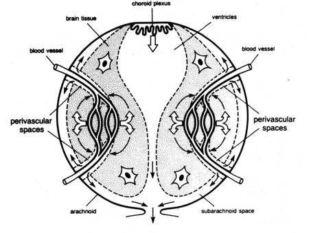
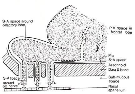
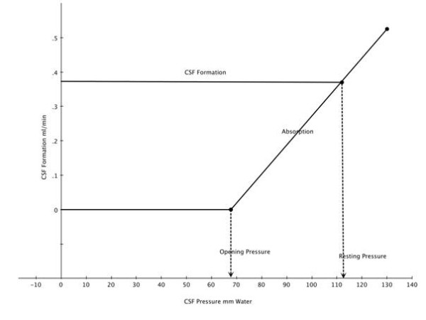
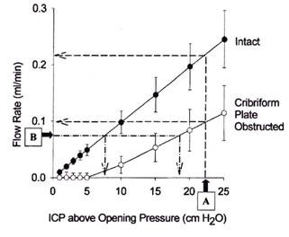
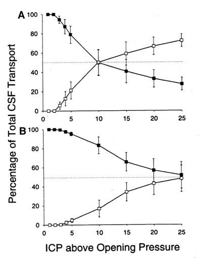
This review traces the development of our understanding of the anatomy and physiological properties of the two systems responsible for the drainage of cerebrospinal fluid (CSF) into the systemic circulation. The roles of the cranial and spinal arachnoid villi (AV) and the lymphatic outflow systems are evaluated as to the dominance of one over the other in various species and degree of animal maturation. The functional capabilities of the total CSF drainage system are presented, with evidence that the duality of the system is supported by the changes in fluid outflow dynamics in human and sub-human primates in hydrocephalus. The review also reconciles the relative importance and alterations of each of the outflow systems in a variety of clinical pathological conditions.


Figures

Figure 1
Figure 1
Experimental arrangement of chamber enclosing arachnoid villi. Upper chamber enclosing sinus side of dura containing arachnoid villi while lower chamber the external surface of the sinus dura (reproduced with permission from Welch and Friedman [5]).
Figure 2
Figure 2
Cerebrospinal fluid valves after reversal of CSF-to-blood pressure gradient. A: Open villus (VO) structure when pressure gradient is positive. B: collapsed villus (VC) when the gradient is negative (reproduced with permission from Welch and Friedman [5]).
Figure 3
Figure 3
Flow-pressure curve through dural disc containing arachnoid villi. Right side: normal flow direction-subarachnoid space to lumen of sagittal sinus. Left side: Flow from sinus to subarachnoid space (reproduced with permission from Welch and Friedman [5]).
Figure 4
Figure 4
Diagrammatic representation of intracellular vacuolation process of cells in canal of Schlemm (reproduced with permission from Tripathi [20]).
Figure 5
Figure 5
Scanning electron micrograph of giant vacuole in mesothelial cell lining of the arachnoid villus seen from the apical aspect with passage of tracer material (colloidal suspended Thorotrast), seen here through the natural opening on the apical surface of the vacuole (arrows; reproduced with permission from Tripathi [19]).
Figure 6
Figure 6
Electron micrograph of the mesothelial cells lining the arachnoid villus showing empty giant vacuole (V). The vacuole on the right has both basal and apical openings, thus constituting a vacuolar transcellular channel (long arrow). The vacuole on the left has basal opening only (short arrows). SAS: subarachnoid space; L: dura lacuna (reproduced with permission from Tripathi [19]).
Figure 7
Figure 7
Diagrammatic representations of the types of meningeal and vascular relationships found in spinal nerve roots. A: arachnoidal cells within dura mater. B: complete penetration of arachnoid villus into interstitium surrounding spinal root. C: penetration of arachnoid villus into epidural spinal vein D and E arachnoidal proliferations within subarachnoid space (reproduced with permission from Welch and Pollay [37]).
Figure 8
Figure 8
Diagrammatic representations of two models of olfactory perineural pathway to nasal lymphatic outflow system (reproduced with permission from Jackson et al [48]).
Figure 9
Figure 9
Schematic diagram illustrating a model of interstitial fluid turnover in the brain based on secretion of cerebral ISF by the blood brain barrier (open arrows) and bulk flow of ISF from brain to CSF via perivascular spaces (curved arrows). CSF is secreted by the choroid plexus (open arrows) and drains with ISF from the subarachnoid space into venous blood and lymph (reproduced with permission from Cserr et al [51]).
Figure 10
Figure 10
Schematic of perineural pathways along cranial nerves for subarachnoid CSF- lymphatic connections (thin curved arrows) and into cranial venous blood via arachnoid villi (large curved arrows; reproduced with permission from Cserr et al [51]).
Figure 11
Figure 11
Schematic of CSF outflow along subarachnoid pathways into nasal lymphatics via olfactory nerve perineural pathway after injection into caudate nucleus (reproduced with permission from Bradbury et al [57]).
Figure 12
Figure 12
Superimposed regression lines for CSF formation and absorption as a function of outflow pressure. The intercept at 112 mm indicates the pressure at which formation and absorption are equal. The pressure at which absorption is zero is also indicated (modified and reproduced with permission from Cutler et al [90]).
Figure 13
Figure 13
Relationship between intracranial pressure (ICP) and flow rate (CSF absorption). CSF access to the spinal subarachnoid compartment was prevented. Closed circles represent data obtained before and open circles represent data obtained after the cribiform plate had been sealed. Opening pressure was the estimated threshold pressure at which CSF absorption was induced (reproduced with permission from Johnston and Papaiconomou [72]).
Figure 14
Figure 14
Estimates of the proportion of total CSF transport through the cribiform plate (solid circles) and other pathways (cranial and spinal arachnoid villi) open circles in constant flow (A) and constant pressure (B) experiments. Cribiform drainage is the dominant location at low and moderate ICP pressures. In A the spinal compartment is intact while in B the spinal compartment is blocked (reproduced with permission from Mollanji et al [100]).

Similar articles

Cited by

References

    1. Brunori A, Vagnozzi R, Giuffre R. Antonio Pacchioni (1665-1726) early studies of the dura mater. J Neurosurg. 1993;78:515–518. doi: 10.3171/jns.1993.78.3.0515. - DOI - PubMed
    1. Trolard D. Les Lacunes Veineuses de la dura-mere. J de L'anatomie. 1892;38:28–56.
    1. Davson H, Segal MB. Physiology of the Cerebrospinal Fluid and Blood-Brain Barriers. London: CRC Press; 1996.
    1. Key A, Retzius G. Studien in der Anatomie des Nervensystems und des Bindegewebes. Stockholm: Samsin and Wallen; 1875.
    1. Welch K, Friedman V. The Cerebrospinal fluid valves. Brain. 1960;83:454–469. doi: 10.1093/brain/83.3.454. - DOI - PubMed

LinkOut - more resources