

Morphological and molecular evidence for functional organization along the rostrocaudal axis of the adult zebrafish intestine
- PMID: 20565988
- PMCID: PMC2996925
- DOI: 10.1186/1471-2164-11-392
Morphological and molecular evidence for functional organization along the rostrocaudal axis of the adult zebrafish intestine
Abstract
Background: The zebrafish intestine is a simple tapered tube that is folded into three sections. However, whether the intestine is functionally similar along its length remains unknown. Thus, a systematic structural and functional characterization of the zebrafish intestine is desirable for future studies of the digestive tract and the intestinal biology and development.
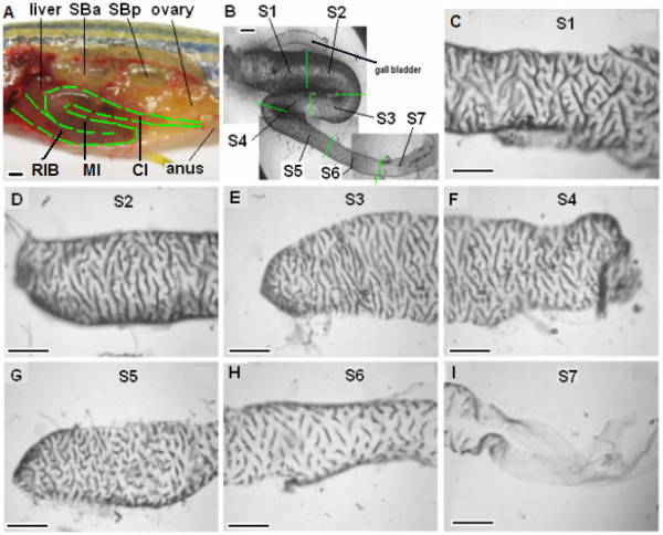
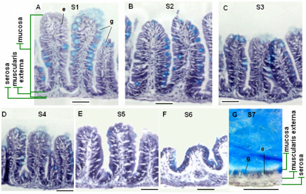
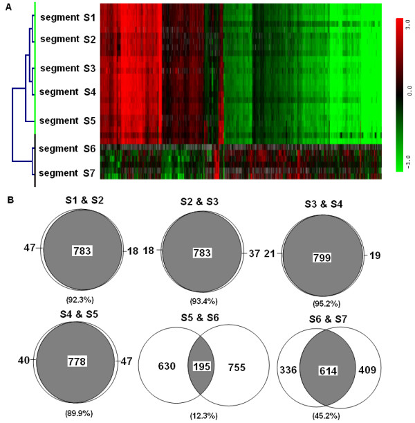
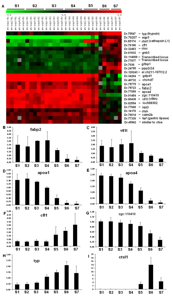
Results: To characterize the structure and function of the adult zebrafish intestine, we divided the intestine into seven roughly equal-length segments, S1-S7, and systematically examined the morphology of the mucosal lining, histology of the epithelium, and molecular signatures from transcriptome analysis. Prominent morphological features are circumferentially-oriented villar ridges in segments S1-S6 and the absence of crypts. Molecular characterization of the transcriptome from each segment shows that segments S1-S5 are very similar while S6 and S7 unique. Gene ontology analyses reveal that S1-S5 express genes whose functions involve metabolism of carbohydrates, transport of lipids and energy generation, while the last two segments display relatively limited function. Based on comparative Gene Set Enrichment Analysis, the first five segments share strong similarity with human and mouse small intestine while S6 shows similarity with human cecum and rectum, and S7 with human rectum. The intestinal tract does not display the anatomical, morphological, and molecular signatures of a stomach and thus we conclude that this organ is absent from the zebrafish digestive system.
Conclusions: Our genome-wide gene expression data indicate that, despite the lack of crypts, the rostral, mid, and caudal portions of the zebrafish intestine have distinct functions analogous to the mammalian small and large intestine, respectively. Organization of ridge structures represents a unique feature of zebrafish intestine, though they produce similar cross sections to mammalian intestines. Evolutionary lack of stomach, crypts, Paneth cells and submucosal glands has shaped the zebrafish intestine into a simpler but unique organ in vertebrate intestinal biology.
Figures
Similar articles
-
Formation of the digestive system in zebrafish: III. Intestinal epithelium morphogenesis.Dev Biol. 2005 Oct 1;286(1):114-35. doi: 10.1016/j.ydbio.2005.07.013. Dev Biol. 2005. PMID: 16125164
-
Mechanical properties of the human gastrointestinal tract.J Biomech. 2002 Oct;35(10):1417-25. doi: 10.1016/s0021-9290(02)00084-2. J Biomech. 2002. PMID: 12231288
-
Regulatory sequences of the mouse villin gene that efficiently drive transgenic expression in immature and differentiated epithelial cells of small and large intestines.J Biol Chem. 1999 Mar 5;274(10):6476-82. doi: 10.1074/jbc.274.10.6476. J Biol Chem. 1999. PMID: 10037740
-
Modeling intestinal disorders using zebrafish.Methods Cell Biol. 2017;138:241-270. doi: 10.1016/bs.mcb.2016.11.006. Epub 2017 Jan 7. Methods Cell Biol. 2017. PMID: 28129846 Review.
-
Gastrointestinal absorption. I. Mechanisms.Am J Pharm Sci Support Public Health. 1975 Sep-Oct;147(5):135-46. Am J Pharm Sci Support Public Health. 1975. PMID: 3972 Review. No abstract available.
Cited by
-
Chronic Exposure to Environmentally Relevant Concentrations of Tetracycline Perturbs Gut Homeostasis in Zebrafish.Environ Health (Wash). 2023 Sep 7;1(4):258-269. doi: 10.1021/envhealth.3c00072. eCollection 2023 Oct 20. Environ Health (Wash). 2023. PMID: 39474494 Free PMC article.
-
Xylanase enhances gut microbiota-derived butyrate to exert immune-protective effects in a histone deacetylase-dependent manner.Microbiome. 2024 Oct 21;12(1):212. doi: 10.1186/s40168-024-01934-6. Microbiome. 2024. PMID: 39434145 Free PMC article.
-
Comprehensive and quantitative proteomic analyses of zebrafish plasma reveals conserved protein profiles between genders and between zebrafish and human.Sci Rep. 2016 Apr 13;6:24329. doi: 10.1038/srep24329. Sci Rep. 2016. PMID: 27071722 Free PMC article.
-
Could a swimming creature inform us on intestinal diseases? Lessons from zebrafish.Inflamm Bowel Dis. 2014 May;20(5):956-66. doi: 10.1097/01.MIB.0000442923.85569.68. Inflamm Bowel Dis. 2014. PMID: 24577115 Free PMC article. Review.
-
Protective mechanisms of a microbial oil against hypercholesterolemia: evidence from a zebrafish model.Front Nutr. 2023 Jun 26;10:1161119. doi: 10.3389/fnut.2023.1161119. eCollection 2023. Front Nutr. 2023. PMID: 37435570 Free PMC article.
References
-
- Lam SH, Wu YL, Vega VB, Miller LD, Spitsbergen J, Tong Y, Zhan H, Govindarajan KR, Lee S, Mathavan S, Murthy KR, Buhler DR, Liu ET, Gong Z. Conservation of gene expression signatures between zebrafish and human liver tumors and tumor progression. Nature Biotechnology. 2006;24:73–75. doi: 10.1038/nbt1169. - DOI - PubMed
Publication types
MeSH terms
LinkOut - more resources
Full Text Sources
Molecular Biology Databases
