. 2007 Mar 15;13 Suppl 1(Suppl 1):123-30.
doi: 10.1177/15910199070130S118. Epub 2007 Jun 27.

Selective transvenous coil embolization of dural arteriovenous fistula. A report of three cases

Affiliations

Selective transvenous coil embolization of dural arteriovenous fistula. A report of three cases

T Yoshioka et al. Interv Neuroradiol. .

Abstract

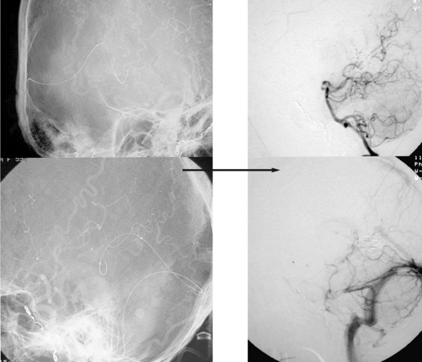
We herein report three cases of dural arteriovenous fistula (DAVF) in which the venous outlet immediately adjacent to the fistula was selectively embolized. Case 1: A 69-year-old man presented with a subarachnoid hemorrhage (SAH). Angiography demonstrated a DAVF in the left superior petrous sinus. Case 2: A 59-yearold woman presented with dizziness. Angiography demonstrated a DAVF adjacent to great vein of Galen. The DAVF drained through the great vein of Galen with retrograde leptomeningeal venous drainage (RLVD). The basal vein of Rosenthal was enhanced from the great vein of Galen. Case 3: A 51-year-old man presented with an occipital seizure. Angiography demonstrated a DAVF adjacent to the left side of the superior sagittal sinus with RLVD. All three cases were successfully treated by the selective embolization of the venous outlet immediately adjacent to the fistula. Therefore, selective embolization preserved normal venous return.


Figures

Figure 1
Figure 1
Case 1. A,C) Left common carotid artery angiogram (early phase). B,D) Left common carotid artery angiogram (late phase). Left common carotid artery angiogram showed a DAVF at the left superior petrous sinus fed by the left occipital artery, left middle meningeal artery and left meningohypophysial trunk. It drained through the left lateral mesencephalic vein and left inferior vermian vein with varicose change.
Figure 2
Figure 2
Case 1. A microcatheter (Terumo) was selectively advanced into the vein and then it was directly connected to the fistula via right jugular vein, right transverse sinus and left inferior vermian vein. The varicose vein connecting to the DAVF was selectively embolized with GDC. After embolization, angiography demonstrated an angiographycal cure without the obliteration of the normal venous return.
Figure 3
Figure 3
Case 2. A) Left vertebral artery angiogram. B) Right common carotid artery angiogram. C) Left common carotid artery angiogram. Angiography demonstrated a DAVF adjacent to great vein of Galen fed by bilateral superficial temporal artery, bilateral middle meningeal artery, bilateral occipital artery, bilateral ascending pharyngeal artery and bilateral tentrial branch of posterior cerebral artery. It drained through the great vein of Galen with retrograde leptomeningeal venous drainage. The basal vein of Rosenthal was enhanced from the great vein of Galen.
Figure 4
Figure 4
Case 2. A microcatheter was selectively advanced to the vein immediately adjacent to the DAVF. The vein immediately adjacent to the DAVF was embolized with GDC. After embolization, angiography demonstrated an angiographycal cure without any obliteration of the normal venous return.
Figure 5
Figure 5
Case 3. Angiography demonstrated a DAVF adjacent to the left side of the superior sagittal sinus. The DAVF was fed by the bilateral superior temporal artery, bilateral middle meningeal artery and left occipital artery. In addition, it also drained through a distended cortical vein connecting to the superior sagittal sinus and retrograde leptomeningeal venous drainage.
Figure 6
Figure 6
Case 3. A) A microcatheter was selectively advanced to the vein immediately adjacent to the DAVF. A varicose vein connecting with DAVF was selectively embolized with GDC. B) A coil mass can be seen embolizing the vein immediately adjacent to the DAVF. C) After embolization, angiography demonstrated an angiographical cure.

Similar articles

Cited by

References

    1. Angiographic predictors of intracranial hemorrhage and clinical outcome in nonsurgical patients. J Neurosurg. 1994;81:531–538. - PubMed
    1. Davis MA, terBrugge K, et al. The validity of classification for the clinical presentation of intracranial arteriovenous fistulas. J Neurosurg. 1996;85:830–837. - PubMed
    1. Lasjaunias P, Chiu M, et al. Neurosurgical manifestations of intracranial arteriovenous malformations. J Neurosurg. 1986;64:724–730. - PubMed
    1. Malik GM, Pearce JE, et al. Dural arteriovenous malformations and intracranial hemorrhage. Neurosurgery. 1984;15:332–339. - PubMed
    1. Colloce M, Dí Aliberti G, et al. Surgical treatment of intracranial dural arteriovenous fistulae: role of venous drainage. Neurosurgery. 2000;47:56–67. - PubMed

LinkOut - more resources