

Patterns of cranial venous system from the comparative anatomy in vertebrates. Part I, introduction and the dorsal venous system
- PMID: 20566102
- PMCID: PMC3329239
- DOI: 10.1177/159101990701300404
Patterns of cranial venous system from the comparative anatomy in vertebrates. Part I, introduction and the dorsal venous system
Abstract
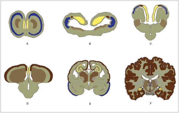
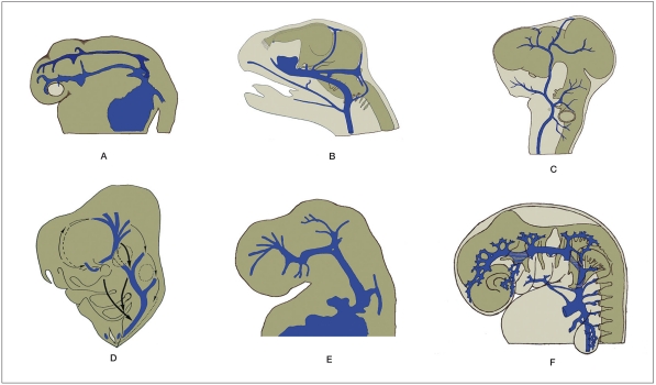
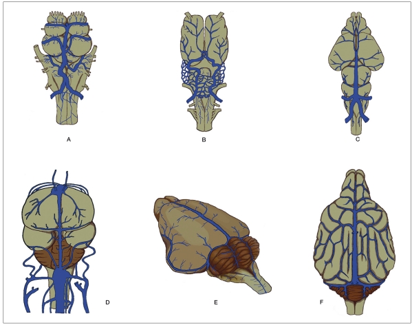
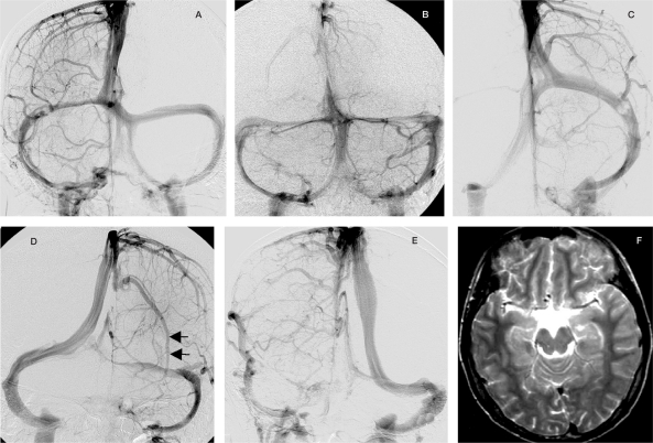
Many classifications of the cerebral venous system are found in the literature but they are seldom based on phylogenic study. Among vertebrates, venous drainage of the brain vesicles differs depending on the species. Due to the variability, poorly descriptive articles, and many different names used for the veins, the comparative study of the cranial venous system can hardly be performed in detail. The cranial venous system in vertebrates can be divided into three systems based on the evolution of the meninges and structures of the brain vesicles: the dorsal, lateral-ventral and ventricular systems. This study proposes a new classification of the venous drainage of brain vesicles using knowledge from a comparative study of vertebrates and focusing on the dorsal venous system. We found that the venous drainage of the neopallium and neocerebellum is involved with this system which may be a recent acquisition of cranial venous evolution.
Figures
Similar articles
-
Patterns of the Cranial Venous System from the Comparative Anatomy in Vertebrates. Part II.The Lateral-Ventral Venous System.Interv Neuroradiol. 2008 Mar 30;14(1):21-31. doi: 10.1177/159101990801400103. Epub 2008 May 12. Interv Neuroradiol. 2008. PMID: 20557782 Free PMC article.
-
Patterns of the Cranial Venous System from the Comparative Anatomy inVertebrates. Part III. The Ventricular System and Comparative Anatomy of the Venous Outlet of Spinal Cord and Its Homology with the Five Brain Vesicles.Interv Neuroradiol. 2008 Jun 30;14(2):125-36. doi: 10.1177/159101990801400203. Epub 2008 Jun 30. Interv Neuroradiol. 2008. PMID: 20557753 Free PMC article.
-
Cranial dural arteriovenous shunts. Part 3. Classification based on the leptomeningeal venous drainage.Neurosurg Rev. 2015 Apr;38(2):273-81; discussion 281. doi: 10.1007/s10143-014-0596-9. Epub 2014 Dec 18. Neurosurg Rev. 2015. PMID: 25516093
-
Cranial dural arteriovenous shunts. Part 1. Anatomy and embryology of the bridging and emissary veins.Neurosurg Rev. 2015 Apr;38(2):253-63; discussion 263-4. doi: 10.1007/s10143-014-0590-2. Epub 2014 Dec 3. Neurosurg Rev. 2015. PMID: 25468011 Review.
-
Anatomy of cerebral veins and sinuses.Front Neurol Neurosci. 2008;23:4-15. doi: 10.1159/000111256. Front Neurol Neurosci. 2008. PMID: 18004050 Review.
Cited by
-
Embryological Consideration of Dural Arteriovenous Fistulas.Neurol Med Chir (Tokyo). 2016 Sep 15;56(9):544-51. doi: 10.2176/nmc.oa.2015-0313. Epub 2016 Jun 1. Neurol Med Chir (Tokyo). 2016. PMID: 27250699 Free PMC article.
-
The median vein of prosencephalon of Markowski: From morphology to genetics.Interv Neuroradiol. 2020 Dec;26(6):752-756. doi: 10.1177/1591019920935316. Epub 2020 Jun 17. Interv Neuroradiol. 2020. PMID: 33283591 Free PMC article. No abstract available.
-
Cerebral Vein Malformations Result from Loss of Twist1 Expression and BMP Signaling from Skull Progenitor Cells and Dura.Dev Cell. 2017 Sep 11;42(5):445-461.e5. doi: 10.1016/j.devcel.2017.07.027. Epub 2017 Aug 30. Dev Cell. 2017. PMID: 28844842 Free PMC article.
-
Group B Streptococcal Neonatal Meningitis.Clin Microbiol Rev. 2022 Apr 20;35(2):e0007921. doi: 10.1128/cmr.00079-21. Epub 2022 Feb 16. Clin Microbiol Rev. 2022. PMID: 35170986 Free PMC article. Review.
-
Derivation of induced pluripotent stem cells from the baboon: a nonhuman primate model for preclinical testing of stem cell therapies.Cell Reprogram. 2013 Dec;15(6):495-502. doi: 10.1089/cell.2012.0093. Epub 2013 Nov 4. Cell Reprogram. 2013. PMID: 24182315 Free PMC article.
References
-
- Jesús Torres-Vázquez MK, Weinstein BM. Molecular distinction between arteries and veins. Cell Tissue Research. 2003;314:43–59. - PubMed
-
- Rhoton ALJ. The cerebral veins. Neurosurgery. 2002;51(4Sup):S159–205. - PubMed
-
- Lasjaunias P, terBrugge KG, Berenstein A, editors. Clinical Vascular anatomy and Variations. 2nd ed. Vol 1. Springer; 2001. Surgical Neuroangiography.
-
- Kent CG, editor. Brain. Comparative Anatomy of the Vertebrates. Times Mirror/Mosby College Publishing; 1987. pp. 530–543.
-
- Montagna W. Comparative anatomy of the central nervous system. Comparative anatomy. New York: John Wiley & Sons, Inc; 1959. pp. 322–339.
LinkOut - more resources
Full Text Sources
