

Arteriovenous fistula of the mandible simulating an odontogenic cyst. A case report
- PMID: 20566108
- PMCID: PMC3329245
- DOI: 10.1177/159101990701300410
Arteriovenous fistula of the mandible simulating an odontogenic cyst. A case report
Abstract
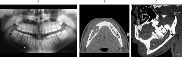
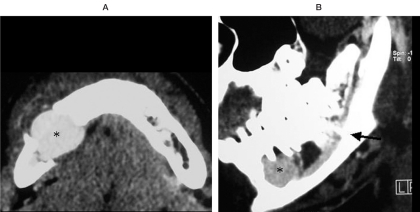
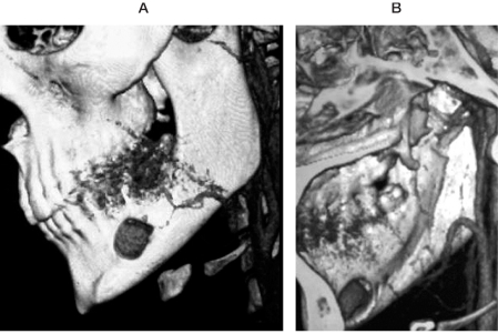
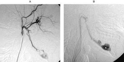
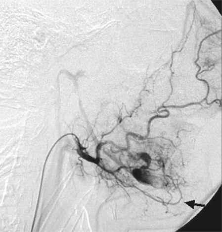
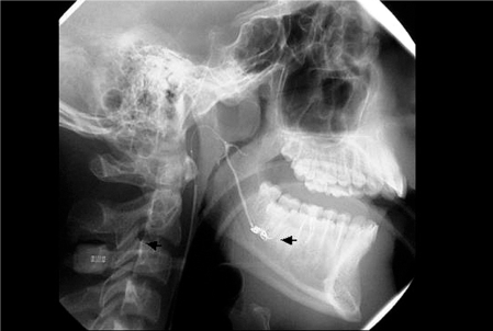
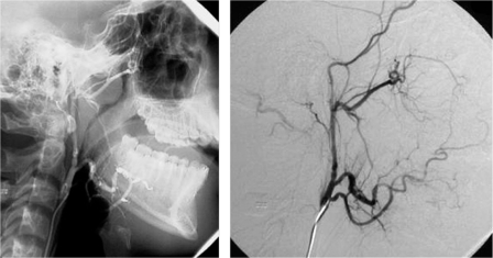
We describe a rare case of mandibular highflow arteriovenous malformation (AVM) mimicking an odontogenic cyst in a young man. The diagnosis of mandibular AVM was made by CT angiography and confirmed by digital subtraction angiography. CT scan showed the extent of mandibular bone alteration and a double enlarged mandibular canal on the same side. An AVM containing a large aneurysm was demonstrated by CT angiography.The mandibular AVM was successfully treated by endovascular therapy with Guglielmi detachable coils. On panoramic radiogram, mandibular AVMs can appear as cystic lesions without pathognomonic features. Several benign and malignant tumours of this anatomical region must be considered in the differential diagnosis.We emphasize the radiological sign of double enlarged mandibular canal and the diagnostic role of CT, particularly CT angiography, to discriminate a mandibular AVM from neoplastic entities of this region, sparing the risks of a needle biopsy.
Figures
Similar articles
-
Arteriovenous fistula of the mandible: a case report.Oral Radiol. 2024 Oct;40(4):555-560. doi: 10.1007/s11282-024-00762-6. Epub 2024 Jun 19. Oral Radiol. 2024. PMID: 38898353 Free PMC article.
-
Mandibular arteriovenous malformation: A rare life-threatening condition depicted on multidetector CT angiography.J Oral Maxillofac Pathol. 2014 Jan;18(1):111-3. doi: 10.4103/0973-029X.131930. J Oral Maxillofac Pathol. 2014. PMID: 24959049 Free PMC article.
-
Treatment of mandibular arteriovenous malformation by transvenous embolization: A case report.Head Neck. 1999 Sep;21(6):574-7. doi: 10.1002/(sici)1097-0347(199909)21:6<574::aid-hed12>3.0.co;2-d. Head Neck. 1999. PMID: 10449675
-
Ruptured Distal Posterior Inferior Cerebellar Artery (PICA) Aneurysms Associated with Cerebellar Arterial Venous Malformations (AVMs): A Case Series and Review of the Literature Demonstrating the Need for Angiographic Evaluation and Feasibility of Endovascular Treatment.World Neurosurg. 2017 Jan;97:751.e7-751.e13. doi: 10.1016/j.wneu.2016.10.081. Epub 2016 Oct 25. World Neurosurg. 2017. PMID: 27793767 Review.
-
Endovascular Management of a Ruptured Basilar Perforator Artery Aneurysm Associated with a Pontine Arteriovenous Malformation: Case Report and Review of the Literature.World Neurosurg. 2018 Aug;116:159-162. doi: 10.1016/j.wneu.2018.05.051. Epub 2018 May 16. World Neurosurg. 2018. PMID: 29777889 Review.
Cited by
-
Arteriovenous fistula of the mandible: a case report.Oral Radiol. 2024 Oct;40(4):555-560. doi: 10.1007/s11282-024-00762-6. Epub 2024 Jun 19. Oral Radiol. 2024. PMID: 38898353 Free PMC article.
References
-
- Lasjaunias P, Berenstein A. Surgical Neuro-angiography. vol 2. Berlin Heidelberg: Springer-Verlag; 1987. Chapter 10 - Craniofacial hemangioma, vascular malformations and angiomatosis: specific aspects; pp. 341–397.
-
- Scholl RJ, Kellett HM, et al. Cysts and cystic lesions of the mandible: clinical and radiologic-histopathologic review. Radiographics. 1999;19:1107–1124. - PubMed
-
- Fan X, Qiu W, et al. Comparative study of clinical manifestations, plain-film radiography, and computed tomographic scan in arteriovenous malformations of the jaws. Oral Surg Oral Med Oral Pathol Oral Radiol Endod. 2002;94:503–509. - PubMed
-
- Mohammadi H, Said-Al-Naief N, Heffez LB. Arteriovenous malformation of the mandible. Oral Surg Oral Med Oral Pathol Oral Radiol Endod. 1997;84:286–289. - PubMed
-
- Rouas P, Nancy J, Bar D. Identification of double mandibular canals: literature review and three case reports with CT-scans and cone beam CT. Dentomaxillofacial Radiology. 2007;36:34–38. - PubMed
LinkOut - more resources
Full Text Sources
