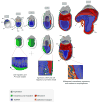
Cellular dynamics in the early mouse embryo: from axis formation to gastrulation
- PMID: 20566281
- PMCID: PMC2908213
- DOI: 10.1016/j.gde.2010.05.008
Cellular dynamics in the early mouse embryo: from axis formation to gastrulation
Abstract
Coordinated cell movements and reciprocal tissue interactions direct the formation of the definitive germ layers and the elaboration of the major axes of the mouse embryo. Genetic and embryological studies have defined the major molecular pathways that mediate these morphogenetic processes and provided 'snapshots' of the morphogenetic program. However, it is increasingly clear that this foundation needs to be validated, and can be significantly refined and extended using live imaging approaches. In situ visualization of these processes in living specimens is a major goal, as it provides unprecedented detail of the individual cellular behaviors, which translate into the large-scale tissue rearrangements that shape the embryo.
Figures


References
-
- Arnold SJ, Robertson EJ. Making a commitment: cell lineage allocation and axis patterning in the early mouse embryo. Nat Rev Mol Cell Biol. 2009;10:91–103. - PubMed
-
Excellent review discussing our current understanding of the molecular mechanisms directing lineage commitment, patterning and morphogensis in the early mouse embryo
-
- Kwon GS, Viotti M, Hadjantonakis AK. The endoderm of the mouse embryo arises by dynamic widespread intercalation of embryonic and extraembryonic lineages. Dev Cell. 2008;15:509–520. - PMC - PubMed
-
Live imaging and genetic labeling are used to investigate the role of the VE during gut endoderm morphogenesis. The study suggests that VE: (1) is dispersed and not diplaced during gut endoderm formation, (2) congregates around midline signaling centers, and (3) derivatives persist in regions of the embryonic gut tube at midgestation
-
- Mesnard D, Guzman-Ayala M, Constam DB. Nodal specifies embryonic visceral endoderm and sustains pluripotent cells in the epiblast before overt axial patterning. Development. 2006;133:2497–2505. - PubMed
-
- Perea-Gomez A, Meilhac SM, Piotrowska-Nitsche K, Gray D, Collignon J, Zernicka-Goetz M. Regionalization of the mouse visceral endoderm as the blastocyst transforms into the egg cylinder. BMC Dev Biol. 2007;7:96. - PMC - PubMed
-
Use of cell labelling, fate mapping and imaging to investigate cell behaviors within the visceral endoderm. Demostration of “polonaise” movements, through retrospective cell arrangement and mixing between emVE and exVE regions
-
- Gibson MC, Patel AB, Nagpal R, Perrimon N. The emergence of geometric order in proliferating metazoan epithelia. Nature. 2006;442:1038–1041. - PubMed
-
This study in Drosophila combines genetics, imaging and mathematical modeling to propose a link between cell geometry and cell behavior in proliferating epithelia
Publication types
MeSH terms
Grants and funding
LinkOut - more resources
Full Text Sources

