Crystal structure of the GalNAc/Gal-specific agglutinin from the phytopathogenic ascomycete Sclerotinia sclerotiorum reveals novel adaptation of a beta-trefoil domain
- PMID: 20566411
- PMCID: PMC2956877
- DOI: 10.1016/j.jmb.2010.05.038
Crystal structure of the GalNAc/Gal-specific agglutinin from the phytopathogenic ascomycete Sclerotinia sclerotiorum reveals novel adaptation of a beta-trefoil domain
Abstract
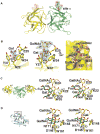
A lectin from the phytopathogenic ascomycete Sclerotinia sclerotiorum that shares only weak sequence similarity with characterized fungal lectins has recently been identified. S. sclerotiorum agglutinin (SSA) is a homodimeric protein consisting of two identical subunits of approximately 17 kDa and displays specificity primarily towards Gal/GalNAc. Glycan array screening indicates that SSA readily interacts with Gal/GalNAc-bearing glycan chains. The crystal structures of SSA in the ligand-free form and in complex with the Gal-beta1,3-GalNAc (T-antigen) disaccharide have been determined at 1.6 and 1.97 A resolution, respectively. SSA adopts a beta-trefoil domain as previously identified for other carbohydrate-binding proteins of the ricin B-like lectin superfamily and accommodates terminal non-reducing galactosyl and N-acetylgalactosaminyl glycans. Unlike other structurally related lectins, SSA contains a single carbohydrate-binding site at site alpha. SSA reveals a novel dimeric assembly markedly dissimilar to those described earlier for ricin-type lectins. The present structure exemplifies the adaptability of the beta-trefoil domain in the evolution of fungal lectins.
Copyright (c) 2010 Elsevier Ltd. All rights reserved.
Figures


Similar articles
-
The Sclerotinia sclerotiorum agglutinin represents a novel family of fungal lectins remotely related to the Clostridium botulinum non-toxin haemagglutinin HA33/A.Glycoconj J. 2007 Apr;24(2-3):143-56. doi: 10.1007/s10719-006-9022-z. Epub 2007 Feb 9. Glycoconj J. 2007. PMID: 17294128
-
Structural and functional characterization of the GalNAc/Gal-specific lectin from the phytopathogenic ascomycete Sclerotinia sclerotiorum (Lib.) de Bary.Biochem Biophys Res Commun. 2003 Aug 22;308(2):396-402. doi: 10.1016/s0006-291x(03)01406-2. Biochem Biophys Res Commun. 2003. PMID: 12901882
-
Structural analysis of the Rhizoctonia solani agglutinin reveals a domain-swapping dimeric assembly.FEBS J. 2013 Apr;280(8):1750-63. doi: 10.1111/febs.12190. Epub 2013 Mar 7. FEBS J. 2013. PMID: 23402398
-
Differential binding properties of Gal/GalNAc specific lectins available for characterization of glycoreceptors.Indian J Biochem Biophys. 1997 Feb-Apr;34(1-2):61-71. Indian J Biochem Biophys. 1997. PMID: 9343930 Review.
-
Differential binding properties of Ga1NAc and/or Ga1 specific lectins.Adv Exp Med Biol. 1988;228:205-63. Adv Exp Med Biol. 1988. PMID: 3051916 Review.
Cited by
-
Agarolytic bacterium Persicobacter sp. CCB-QB2 exhibited a diauxic growth involving galactose utilization pathway.Microbiologyopen. 2017 Feb;6(1):e00405. doi: 10.1002/mbo3.405. Epub 2016 Dec 17. Microbiologyopen. 2017. PMID: 27987272 Free PMC article.
-
Structure- and context-based analysis of the GxGYxYP family reveals a new putative class of glycoside hydrolase.BMC Bioinformatics. 2014 Jun 17;15:196. doi: 10.1186/1471-2105-15-196. BMC Bioinformatics. 2014. PMID: 24938123 Free PMC article.
-
Crystal Structure of Crataeva tapia Bark Protein (CrataBL) and Its Effect in Human Prostate Cancer Cell Lines.PLoS One. 2013 Jun 18;8(6):e64426. doi: 10.1371/journal.pone.0064426. Print 2013. PLoS One. 2013. PMID: 23823708 Free PMC article.
-
Promiscuity of the euonymus carbohydrate-binding domain.Biomolecules. 2012 Oct 8;2(4):415-34. doi: 10.3390/biom2040415. Biomolecules. 2012. PMID: 24970144 Free PMC article.
-
Hitting the sweet spot-glycans as targets of fungal defense effector proteins.Molecules. 2015 May 6;20(5):8144-67. doi: 10.3390/molecules20058144. Molecules. 2015. PMID: 25955890 Free PMC article. Review.
References
-
- Wang H, Ng TB, Ooi VEC. Lectins from mushrooms. Mycological Research. 1998;102:897–906.
-
- Imberty A, Mitchell EP, Wimmerová M. Structural basis of high-affinity glycan recognition by bacterial and fungal lectins. Curr Opin Struct Biol. 2005;15:525–534. - PubMed
-
- Goldstein IJ, Winter . Comprehensive glycoscience: From Chemistry to Systems Biology. Elsevier Ltd; UK: 2007. Mushroom lectins.
-
- Kellens JTC, Peumans WJ. Developmental accumulation of lectins in Rhizoctonia solanii: potential role as a storage protein. J Gen Microbiol. 1990;136:2489–2495.
-
- Rosen S, Sjollema K, Veenhuis M, Tunlid A. A cytoplasmic lectin produced by the fungus Arthrobotrys oligospora functions as a storage protein during saprophytic and parasitic growth. Microbiology. 1997;143:2593. - PubMed
Publication types
MeSH terms
Substances
Associated data
- Actions
- Actions
Grants and funding
LinkOut - more resources
Full Text Sources
Research Materials

