. 2010 Oct;38(19):6490-501.
doi: 10.1093/nar/gkq552. Epub 2010 Jun 21.

The ribonucleotide reductase inhibitor, Sml1, is sequentially phosphorylated, ubiquitylated and degraded in response to DNA damage

Affiliations

The ribonucleotide reductase inhibitor, Sml1, is sequentially phosphorylated, ubiquitylated and degraded in response to DNA damage

Bethany L Andreson et al. Nucleic Acids Res. 2010 Oct.

Abstract

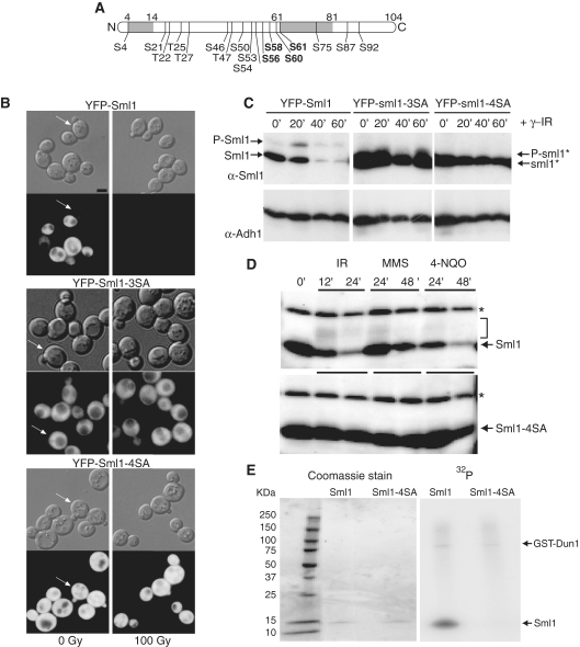
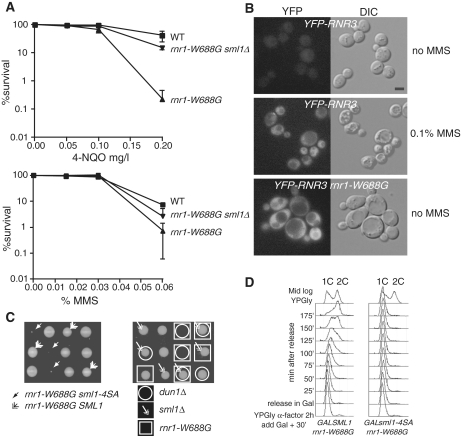
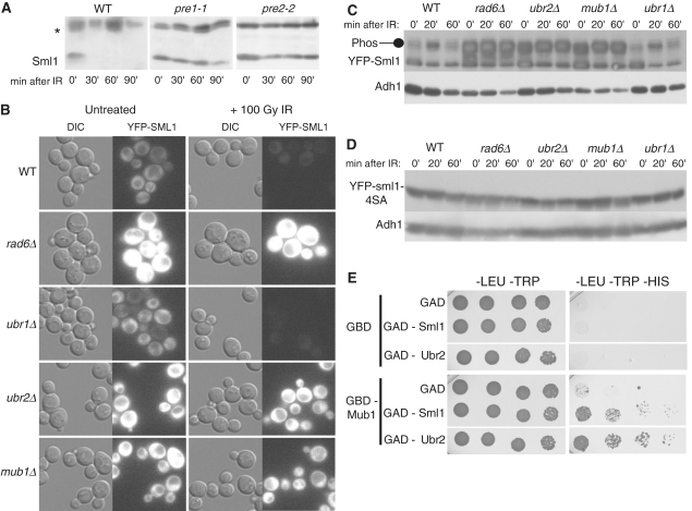
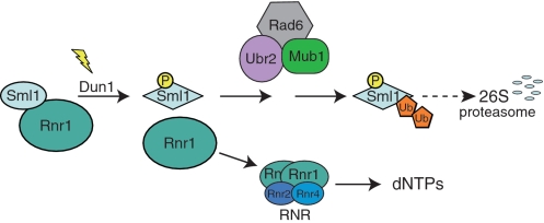
Regulation of ribonucleotide reductase (RNR) is important for cell survival and genome integrity in the face of genotoxic stress. The Mec1/Rad53/Dun1 DNA damage response kinase cascade exhibits multifaceted controls over RNR activity including the regulation of the RNR inhibitor, Sml1. After DNA damage, Sml1 is degraded leading to the up-regulation of dNTP pools by RNR. Here, we probe the requirements for Sml1 degradation and identify several sites required for in vivo phosphorylation and degradation of Sml1 in response to DNA damage. Further, in a strain containing a mutation in Rnr1, rnr1-W688G, mutation of these sites in Sml1 causes lethality. Degradation of Sml1 is dependent on the 26S proteasome. We also show that degradation of phosphorylated Sml1 is dependent on the E2 ubiquitin-conjugating enzyme, Rad6, the E3 ubiquitin ligase, Ubr2, and the E2/E3-interacting protein, Mub1, which form a complex previously only implicated in the ubiquitylation of Rpn4.


Figures

Figure 1.
Figure 1.
Mutations that eliminate putative phosphorylation sites in Sml1 stabilize the protein after DNA damage treatment and prevent in vivo and in vitro phosphorylation. (A) Depiction of the Sml1 protein with the positions of all serine and threonine residues that are potential phosphorylation sites. The shaded regions indicate the positions of two α helices, including the C-terminal helix, which is important for Sml1 binding to Rnr1. The four serines changed in the sml1-4SA mutant are shown in bold type. (B) Mid-log phase cultures of cells expressing YFP-Sml1 (W4622-14B), YFP-sml1-3SA (W6976-4A) or YFP-sml1-4SA (W4748-4D) fusion proteins were treated with 100 Gy of γ-irradiation. Protein stability was examined by visualizing YFP fluorescence before and after treatment. White arrows indicate cells that are in S phase (small buds). The scale bar is equal to 3 µm. (C) Total yeast extracts of the strains shown in (B) were probed with anti-Sml1 antibody to examine stability and in vivo phosphorylation, as determined by mobility shift of immunoblot, of the fusion proteins in logarithmically growing cultures. To control for loading, the membrane was stripped and re-probed using anti-Adh1 antibody. (D) Total yeast extracts from wild-type (W1588-4C) and sml1-4SA (W3329-7D) strains were examined for Sml1 in vivo phosphorylation in response to treatment with γ-irradiation (300 Gy), 0.05% MMS and 4-NQO (0.25 mg/l). The arrow indicates the position of Sml1 proteins. The slower migrating bands (indicated by a bracket) are due to phosphorylation (21). Immunoblots were probed with anti-Sml1 serum. The top band, labeled with an asterisk, is a Sml1-independent cross-reacting band used as a loading control. (E) Recombinant purified Sml1 and sml1-4SA were incubated with GST-Dun1 fusion protein purified from yeast. A portion of the reaction was resolved on a 4–20% SDS–PAGE gradient gel, stained with Coomassie blue and subsequently visualized by autoradiography for 32P incorporation. Both reactions contained the same amount of recombinant protein (left) and exhibited the same level of kinase activity as observed by GST-Dun1 autophosphorylation (right).
Figure 2.
Figure 2.
Overexpression of sml1-4SA, but not SML1, slows S phase progression in wild-type cells. (A) Wild-type and sml1-4SA strains were analyzed for cell-cycle progression. There is no significant difference in the duration of S phase between the two strains when the proteins are expressed endogenously (black lines to the right of the panels). (B) Overexpression of sml1-4SA (W3332-5C), but not SML1 (W2056-8A), driven by a strong galactose promoter slows S phase progression in wild-type cells. Black lines to the right of the panels indicate S-phase.
Figure 3.
Figure 3.
RNR regulation and checkpoint activation are normal in a sml1-4SA strain. (A) Cells expressing YFP-Sml1 (W5530-6C) or YFP-sml1-4SA (W5755-2A) along with CFP-Rnr1 were visualized by fluorescent microscopy before and an hour after 200 Gy of γ-irradiation. Live cell images were captured identically and YFP levels are depicted on the graphs. Cells without buds are G1 cells and were used for subsequent analyses. (B) Protein extracts were taken from wild-type (W1588-4C) and sml1-4SA (W3329-7D) cells before and an hour after 0.03% MMS treatment and analyzed by immunoblot using a Rad53 antibody. (C) Cells expressing both YFP-sml1-4SA and CFP-Rnr2 proteins (W5766-1D) were visualized by fluorescent microscopy before and an hour after 200 Gy of γ-irradiation. WT, wild-type strain.
Figure 4.
Figure 4.
sml1-4SA sensitizes cells that are defective in dNTP regulation. (A) rnr1-W688G is sensitive to genotoxic stress. The DNA damage sensitivities of the strains [wild type (W1588-4C), rnr1-W688G (W4383-1B) and rnr1-W688G sml1Δ (W4383-10C)] were examined in a quantitative survival assay used to determine the LD50 values after 4-NQO (mg/l) and MMS (%) treatments. Mid-log phase cultures were sonicated and appropriate dilutions were plated on YPD for viability, or on YPD plates containing 4NQO (top) and MMS (bottom) at various concentrations. The reported value is the mean of three experiments and the error bars represent the standard error of the mean. (B) rnr1-W688G induces Rnr3 expression in the absence of exogenous damage. The activation of the DNA damage checkpoint was monitored through the induction of a YFP-Rnr3 fusion protein (W6986-1B). Rnr3 is induced in response to DNA damage and the induction depends on checkpoint signaling (55). In the absence of DNA damage, YFP-Rnr3 is not expressed in wild-type cells (top). To induce YFP-Rnr3 expression, mid-log phase cells were treated with 0.1% MMS for 5 h (middle). The rnr1-W688G mutation causes YFP-Rnr3 to be expressed without any exogenous DNA damage treatment (bottom). (C) Dissection of heterozygous diploid SML1/sml1-4SA rnr1-W688G/RNR1 (W4383) and SML1/sml1Δ DUN1/dun1Δ RNR1/rnr1-W688G (W4384) strains shows a genetic interaction between rnr1-W688G and the sml1-4SA and dun1Δ alleles. Deletion of sml1Δ suppresses the synthetic lethality between rnr1-W688G and dun1Δ. The four spores of each tetrad are positioned in the three rows shown. (D) Yeast cells (W3755-14D: GAL-SML1 rnr1-W688G and W3756-3B: GAL-sml1-4SA rnr1-W688G) growing in YPGly at 30°C were synchronized at G1 with α-factor for 2 h. Galactose was added 30 min into the treatment. The pheromone was removed through rapid filtration and cells were released in fresh YPGal without α-factor. At each time point, samples were fixed in 70% ethanol for DNA content analysis by FACS. WT, wild-type strain.
Figure 5.
Figure 5.
Sml1 is degraded by the 26S proteasome and the degradation is dependent on Rad6, Ubr2 and Mub1. (A) Wild-type (MHY686) or mutant cells were shifted to 37°C for 3 h to inactivate the proteasome in the pre1-1 (MHY687) and pre2-2 (MHY689) mutants, and treated with 0.03% MMS. Samples were run on a 15% SDS–PAGE gel. Using anti-Sml1 serum, total yeast extracts were examined for Sml1 protein levels at the indicated time points after MMS treatment. In both immunoblots, the top band, labeled with an asterisk, is a Sml1-independent cross-reacting band used as a loading control. (B) Mid-log cultures of WT, rad6Δ, ubr1Δ, ubr2Δ and mub1 strains containing a genomic copy of YFP-Sml1 (W9174-5D, W9174-10C, W9177-8B, W9175-7C and W9176-4D, respectively) were treated with 100 Gy of γ-irradiation. Protein stability was examined by visualizing YFP fluorescence before and after treatment. (C) (Top panel) Total yeast extracts from logarithmically growing cultures of the strains shown in (B) were immunoblotted and probed with anti-Sml1 antibody to examine stability and in vivo phosphorylation. To control for loading, the membrane was stripped and re-probed using anti-Adh1 antibody. (D) Total yeast extracts of mid-log cultures of WT, rad6Δ, ubr1Δ, ubr2Δ and mub1 strains containing a genomic copy of YFP-sml1-4SA (W9261-7D, W9261-11B, W9264-11C, W9262-2D and W9263-7B, respectively) were treated with 100 Gy of γ-irradiation, immunoblotted and probed with anti-Sml1 antibody. To control for loading, the membrane was stripped and re-probed using anti-Adh1 antibody. (E) Diploids containing different combinations of GBD or GBD-Mub1 with GAD, GAD-Sml1 or GAD-Ubr2 (as indicated) were spotted in 5-fold serial dilutions onto -LEU–TRP and -LEU–TRP–HIS media. The -LEU–TRP medium selects for diploids containing a GBD and GAD plasmids. Growth on medium lacking histidine indicates a two-hybrid interaction and was observed for GBD-Mub1 and GAD-Sml1 as well as GBD-Mub1 and GAD-Ubr2. Plates were scanned after 4 days of growth. WT, wild-type strain.
Figure 6.
Figure 6.
A model for Sml1 regulation in response to DNA damage. Prior to modification, Sml1 is bound to Rnr1 and inhibits RNR activity. Following DNA damage, Dun1 is activated and phosphorylates Sml1 on serines 56, 58, 60 and/or 61. Phosphorylation promotes ubiquitylation of Sml1 by the Rad6–Ubr2–Mub1 complex, targeting Sml1 for degradation by the 26S proteasome. The released Rnr1 associates with Rnr2 and Rnr4 to form an active RNR enzyme allowing the production of dNTPs.

References

    1. Chabes A, Georgieva B, Domkin V, Zhao X, Rothstein R, Thelander L. Survival of DNA damage in yeast directly depends on increased dNTP levels allowed by relaxed feedback inhibition of ribonucleotide reductase. Cell. 2003;112:391–401. - PubMed
    1. Chabes A, Stillman B. Constitutively high dNTP concentration inhibits cell cycle progression and the DNA damage checkpoint in yeast Saccharomyces cerevisiae. Proc. Natl Acad. Sci. USA. 2007;104:1183–1188. - PMC - PubMed
    1. Reichard P. Interactions between deoxyribonucleotide and DNA synthesis. Annu. Rev. Biochem. 1988;57:349–374. - PubMed
    1. Elledge SJ, Davis RW. Two genes differentially regulated in the cell cycle and by DNA-damaging agents encode alternative regulatory subunits of ribonucleotide reductase. Genes Dev. 1990;4:740–751. - PubMed
    1. Elledge SJ, Davis RW. Identification and isolation of the gene encoding the small subunit of ribonucleotide reductase from Saccharomyces cerevisiae: DNA damage-inducible gene required for mitotic viability. Mol. Cell. Biol. 1987;7:2783–2793. - PMC - PubMed

Publication types

MeSH terms

Substances