

p27 suppresses arsenite-induced Hsp27/Hsp70 expression through inhibiting JNK2/c-Jun- and HSF-1-dependent pathways
- PMID: 20566634
- PMCID: PMC2924005
- DOI: 10.1074/jbc.M110.100271
p27 suppresses arsenite-induced Hsp27/Hsp70 expression through inhibiting JNK2/c-Jun- and HSF-1-dependent pathways
Abstract
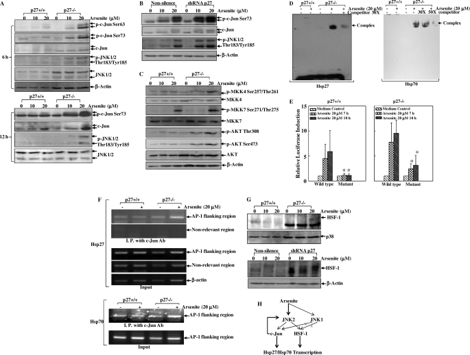
p27 is an atypical tumor suppressor that can regulate the activity of cyclin-dependent kinases and G(0)-to-S phase transitions. More recent studies reveal that p27 may also exhibit its tumor-suppressive function through regulating many other essential cellular events. However, the molecular mechanisms underlying these anticancer effects of p27 are largely unknown. In this study, we found that depletion of p27 expression by either gene knock-out or knockdown approaches resulted in up-regulation of both Hsp27 and Hsp70 expression at mRNA- and promoter-derived transcription as well as protein levels upon arsenite exposure, indicating that p27 provides a negative signal for regulating the expression of Hsp27 and Hsp70. Consistently, arsenite-induced activation of JNK2/c-Jun and HSF-1 pathways was also markedly elevated in p27 knock-out (p27(-/-)) and knockdown (p27 shRNA) cells. Moreover, interference with the expression or function of JNK2, c-Jun, and HSF-1, but not JNK1, led to dramatic inhibition of arsenite-induced Hsp27 and Hsp70 expression. Collectively, our results demonstrate that p27 suppresses Hsp27 and Hsp70 expression at the transcriptional level specifically through JNK2/c-Jun- and HSF-1-dependent pathways upon arsenite exposure, which provides additional important molecular mechanisms for the tumor-suppressive function of p27.
Figures


Similar articles
-
p27 suppresses cyclooxygenase-2 expression by inhibiting p38β and p38δ-mediated CREB phosphorylation upon arsenite exposure.Biochim Biophys Acta. 2013 Sep;1833(9):2083-91. doi: 10.1016/j.bbamcr.2013.04.012. Epub 2013 Apr 29. Biochim Biophys Acta. 2013. PMID: 23639288 Free PMC article.
-
Arsenite stabilizes HIF-1α protein through p85α-mediated up-regulation of inducible Hsp70 protein expression.Cell Mol Life Sci. 2011 Feb;68(3):475-88. doi: 10.1007/s00018-010-0459-7. Epub 2010 Sep 12. Cell Mol Life Sci. 2011. PMID: 20835880 Free PMC article.
-
Prostaglandins stimulate the stress-induced synthesis of hsp27 and alpha B crystallin.J Cell Physiol. 1997 Mar;170(3):255-62. doi: 10.1002/(SICI)1097-4652(199703)170:3<255::AID-JCP6>3.0.CO;2-N. J Cell Physiol. 1997. PMID: 9066782
-
Coordination of JNK1 and JNK2 is critical for GADD45alpha induction and its mediated cell apoptosis in arsenite responses.J Biol Chem. 2006 Nov 10;281(45):34113-23. doi: 10.1074/jbc.M602821200. Epub 2006 Sep 13. J Biol Chem. 2006. PMID: 16973625
-
HSP27 regulates p53 transcriptional activity in doxorubicin-treated fibroblasts and cardiac H9c2 cells: p21 upregulation and G2/M phase cell cycle arrest.Am J Physiol Heart Circ Physiol. 2008 Apr;294(4):H1736-44. doi: 10.1152/ajpheart.91507.2007. Epub 2008 Feb 8. Am J Physiol Heart Circ Physiol. 2008. Retraction in: Am J Physiol Heart Circ Physiol. 2024 Aug 1;327(2):H445. doi: 10.1152/ajpheart.91507.2007_RET. PMID: 18263706 Retracted.
Cited by
-
Functional HSF1 requires aromatic-participant interactions in protecting mouse embryonic fibroblasts against apoptosis via G2 cell cycle arrest.Mol Cells. 2012 May;33(5):465-70. doi: 10.1007/s10059-012-2246-9. Epub 2012 Apr 17. Mol Cells. 2012. PMID: 22526392 Free PMC article.
-
Selenium-binding protein 1 transcriptionally activates p21 expression via p53-independent mechanism and its frequent reduction associates with poor prognosis in bladder cancer.J Transl Med. 2020 Jan 9;18(1):17. doi: 10.1186/s12967-020-02211-4. J Transl Med. 2020. PMID: 31918717 Free PMC article.
-
Isorhapontigenin (ISO) inhibited cell transformation by inducing G0/G1 phase arrest via increasing MKP-1 mRNA Stability.Oncotarget. 2014 May 15;5(9):2664-77. doi: 10.18632/oncotarget.1872. Oncotarget. 2014. PMID: 24797581 Free PMC article.
-
p27 suppresses cyclooxygenase-2 expression by inhibiting p38β and p38δ-mediated CREB phosphorylation upon arsenite exposure.Biochim Biophys Acta. 2013 Sep;1833(9):2083-91. doi: 10.1016/j.bbamcr.2013.04.012. Epub 2013 Apr 29. Biochim Biophys Acta. 2013. PMID: 23639288 Free PMC article.
-
p63α protein up-regulates heat shock protein 70 expression via E2F1 transcription factor 1, promoting Wasf3/Wave3/MMP9 signaling and bladder cancer invasion.J Biol Chem. 2017 Sep 22;292(38):15952-15963. doi: 10.1074/jbc.M117.792010. Epub 2017 Aug 9. J Biol Chem. 2017. PMID: 28794159 Free PMC article.
References
-
- Kim D., Kim S. H., Li G. C. (1999) Biochem. Biophys. Res. Commun. 254, 264–268 - PubMed
-
- Garrido C., Gurbuxani S., Ravagnan L., Kroemer G. (2001) Biochem. Biophys. Res. Commun. 286, 433–442 - PubMed
-
- Garrido C., Schmitt E., Candé C., Vahsen N., Parcellier A., Kroemer G. (2003) Cell Cycle 2, 579–584 - PubMed
-
- Sgambato A., Cittadini A., Faraglia B., Weinstein I. B. (2000) J. Cell. Physiol. 183, 18–27 - PubMed
-
- Wang J. P., Qi L., Moore M. R., Ng J. C. (2002) Toxicol. Lett. 133, 17–31 - PubMed
Publication types
MeSH terms
Substances
Grants and funding
LinkOut - more resources
Full Text Sources
Research Materials
Miscellaneous
