. 2010 Aug 20;285(34):26358-68.
doi: 10.1074/jbc.M110.147439. Epub 2010 Jun 21.

Cystathionine gamma-Lyase-deficient mice require dietary cysteine to protect against acute lethal myopathy and oxidative injury

Affiliations

Cystathionine gamma-Lyase-deficient mice require dietary cysteine to protect against acute lethal myopathy and oxidative injury

Isao Ishii et al. J Biol Chem. .

Abstract

Cysteine is considered a nonessential amino acid in mammals as it is synthesized from methionine via trans-sulfuration. However, premature infants or patients with hepatic failure may require dietary cysteine due to a lack of cystathionine gamma-lyase (CTH), a key trans-sulfuration enzyme. Here, we generated CTH-deficient (Cth(-/-)) mice as an animal model of cystathioninemia/cystathioninuria. Cth(-/-) mice developed normally in general but displayed hypercystathioninemia/hyperhomocysteinemia though not hypermethioninemia. When fed a low cyst(e)ine diet, Cth(-/-) mice showed acute skeletal muscle atrophy (myopathy) accompanied by enhanced gene expression of asparagine synthetase and reduced contents of glutathione in livers and skeletal muscles, and intracellular accumulation of LC3 and p62 in skeletal myofibers; they finally died of severe paralysis of the extremities. Cth(-/-) hepatocytes required cystine in a culture medium and showed greater sensitivity to oxidative stress. Cth(-/-) mice exhibited systemic vulnerability to oxidative injury, which became more prominent when they were fed the low cyst(e)ine diet. These results reveal novel roles of trans-sulfuration previously unrecognized in mice lacking another trans-sulfuration enzyme cystathionine beta-synthase (Cbs(-/-)). Because Cbs(-/-) mice display hyperhomocysteinemia and hypermethioninemia, our results raise questions against the homocysteine-based etiology of CBS deficiency and the current newborn screening for homocysteinemia using Guthrie's method, which detects hypermethioninemia.


Figures
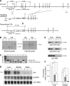
FIGURE 1.
FIGURE 1.
Targeted deletion of CTH in mice. A, maps of the WT Cth locus including a total of 12 exons that encode the full ORF, the targeting construct, and the homologously recombined locus. Approximate positions for two Southern probes and three PCR primers are indicated. B, BamH I; pBS, pBluescript; DTA, diphtheria toxin A gene; LacZ, β-galactosidase gene; Neo, neomycin phosphotransferase gene. B, Southern blot analysis of BamH I-digested genomic DNA isolated from WT/recombined embryonic stem cells and tails of Cth mutant mice. Blots were hybridized with either an external or internal probe. C, PCR genotyping using tail DNA and three PCR primers (primers 1–3) detecting the inheritance of WT and recombined Cth alleles (299- and 194-bp, respectively). D, Northern blot analysis of RNA from 8-week-old male livers, kidneys, and brains using the full-length Cth probe. GAPDH blots and ethidium bromide-stained 18 S rRNA are loading controls. E, Western blot analysis of 8-week-old male liver/kidney homogenates using polyclonal antibodies raised against a recombinant CTH amino terminus (N-ter), CTH carboxyl terminus (C-ter), and full-length CBS. F, specific CTH activity in 8-week-old male liver/kidney homogenate. Values are mean ± S.D. (n = 3); *, p < 0.01 and ‡, p < 0.001 in the one-way ANOVA.
FIGURE 2.
FIGURE 2.
Low cyst(e)ine diet-induced acute muscular atrophy in Cth−/− mice. A, body weight changes in WT (blue circles), Cth+/− (green triangles), and Cth−/− (red diamonds) females that were fed the KR diet after weaning at 3 weeks of age. All Cth−/− mice died after ∼2 weeks. B and C, the appearance (B) and a radiograph (C) of WT (left) and Cth−/− (right) females fed the KR diet for 1 week from 3 weeks of age. Severe atrophy was observed in abdominal regions (B) as well as in proximal muscles such as trapezius and rectus femoris muscles (arrow and arrowhead in C, respectively). Bars, 1 cm. D, body weight changes with the KR+Cys diet. E, hematoxylin/eosin-stained transverse cross-sections of rectus femoris muscles isolated from a WT or Cth−/− male fed the KR diet for 1 week from 3 weeks of age. Diffuse muscular atrophy was observed in Cth−/− mice. Bars, 50 μm. F, myofiber cross-sectional areas (μm2) of rectus femoris muscles from WT or Cth−/− males before (3 weeks of age) and after (4 weeks of age) 1 week on the KR diet. G, serum levels of creatinine after consumption of the standard (Std) or KR diet for 1 week from 3 weeks of age. H and I, body weight changes after a change from the standard diet to KR diet at 6 (H) and 12 (I) weeks of age. After the change, both 6- and 12-week-old Cth−/− lost weight daily and died within ∼4 weeks. J and K, the daily body weight change (J) and food intake (K) in WT or Cth−/− males after the change to the KR diet at 8 weeks of age. Representative pictures are shown in B, C, and E. Values are mean ± S.D. (n = 5 for each genotype in A, D, H, and I; 6 in F; 10 in G; and 8 in J and K). *, p < 0.05; ‡, p < 0.01; and †, p < 0.001 in the one-way ANOVA in F and G; and ‡, p < 0.01 in the t test in J. The asterisks for p values are abbreviated in A, H, and I.
FIGURE 3.
FIGURE 3.
Increased expression of asparagine synthetase gene in the liver and muscle of KR-fed Cth−/− mice. The expression of asparagine synthetase (Asns) in A, livers, B, skeletal muscles, and C, hearts of 9-week-old WT and Cth−/− males that were fed the standard (Std) diet, the KR diet (for 1 week) or starved (Starv) for 2 days, was analyzed by RT-PCR and normalized to that of housekeeping hypoxanthine guanine phosphoribosyl transferase 1 (Hprt1). The Asns/Hprt1 expression ratios in the standard diet-fed WT were set at 1. Values are mean ± S.D. (n = 6). *, p < 0.05; ‡, p < 0.01; and †, p < 0.001 in the t test.
FIGURE 4.
FIGURE 4.
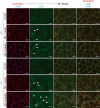
Accumulation of LC3 and p62 in the myofibers of KR-fed Cth−/− mice. Eight-week-old WT and Cth−/− females were fed without or with KR for 1 or 3 weeks, and femur skeletal muscle sections were stained with DAPI, and anti-LC3, dystrophin, or p62 antibodies. LC3-positive punctation appeared in the peripheral regions of both WT and Cth−/− myofibers after 1 week on the KR diet (arrowheads), and LC3 was diffusely increased within some Cth−/− myofibers after 3 weeks of KR feeding (arrows). Diffuse p62 expression was observed in some Cth−/− myofibers after 3 weeks (asterisks). The structure of dystrophin was preserved even in p62-positive myofibers of KR-fed Cth−/− mice. Bars, 25 μm.
FIGURE 5.
FIGURE 5.
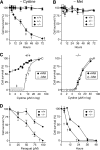
Increased sensitivity of Cth−/− hepatocytes to cystine depletion and oxidative injury. A and B, hepatocytes isolated from 8-week-old WT (circles), Cth+/− (triangles), and Cth−/− (diamonds) males, were cultured in A, cystine- or B, Met-free DMEM supplemented with 10% dialyzed FBS. Cell survivability was evaluated by measuring cellular ATP levels after the indicated hours of incubation. Survivability at 0 h was set as 100%. C, WT (circles in left panel) and Cth−/− (diamonds in right panel) hepatocytes were cultured for 5 days in 0.2 mm Met-containing (closed symbols) or Met-free (open symbols) medium containing varied concentrations of cystine and 10% dialyzed FBS. Met supplementation did not rescue cystine-depleted hepatocytes in the absence of CTH. D, WT and Cth−/− hepatocytes were treated with paraquat. Concentration- (in a 1-day incubation) and time-dependent (at 100 μm) inhibitory effects on cell survival are shown on the left and right, respectively. Values are mean ± S.D. (n = 4); *, p < 0.05; ‡, p < 0.01; and †, p < 0.001 versus +/+ samples (in A, B, and D) or +Met samples (in C) in the t test.
FIGURE 6.
FIGURE 6.
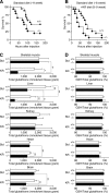
Increased sensitivity of Cth−/− mice to oxidative injury from paraquat and the impact of 1 week on the KR diet on sensitivity and tissue glutathione contents. A, Kaplan-Meier survival analysis after the intraperitoneal injection of paraquat (50 mg/kg) into 9-week-old WT (n = 17) and Cth−/− (n = 15) males. Health status was monitored every 8 h for a week. Cth−/− mice showed higher sensitivity to paraquat than WT (p = 0.038). B, the same as in A except that WT and Cth−/− males (n = 15 each) were fed KR for a week before the injection. KR increased the sensitivity to paraquat in both WT and Cth−/− mice (p = 0.017 and 0.049, respectively). When fed KR for a week, Cth−/− mice still showed greater sensitivity to paraquat than WT (p = 0.031). C, total glutathione (GSH+oxidized glutathione) levels. D, percentages of GSH/total glutathione in skeletal muscle, liver, kidney, heart, and brain extracts in 3-week-old WT (open bars) and Cth−/− (filled bars) females. After 1 week on the KR diet, total glutathione levels decreased in most tissues of WT and Cth−/− and especially Cth−/− mice. Values are mean ± S.D. (n = 5); *, p < 0.05; ‡, p < 0.01; and †, p < 0.001 in the one-way ANOVA. Std, standard.
FIGURE 7.
FIGURE 7.
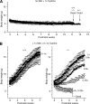
Cth−/− mice require dietary cystine as an essential amino acid. A, after weaning at 3 weeks of age, WT (circles; n = 4), Cth+/− (triangles; n = 9), and Cth−/− (diamonds; n = 4) males were fed a protein-free amino acid diet that contained cystine (0.37%: an amount equivalent to the standard diet) but not Met. No developmental body weight increase was observed in any of the mice up until their death after 8–9 weeks. B, WT (left) and Cth−/− (right) males were fed a protein-free amino acid diet that contained Met (0.44%: an amount equivalent to the standard diet) and an amount of cystine (open circles, 0× (0%); open triangles, 0.23× (0.0851%); open diamonds, 0.45× (0.1665%); filled circles, 0.6× (0.222%); filled triangles, 0.8× (0.296%); and filled diamonds, 1× (0.37%)). Cth−/− but not WT mice required cystine as an essential amino acid. Values are mean ± S.D. (n = 5); the asterisks for p values are abbreviated in B (right panel).

Similar articles

Cited by

References

    1. Finkelstein J. D. (2000) Semin. Thromb. Hemost. 26, 219–225 - PubMed
    1. Stipanuk M. H. (2004) Annu. Rev. Nutr. 24, 539–577 - PubMed
    1. Sturman J. A., Gaull G., Raiha N. C. (1970) Science 169, 74–76 - PubMed
    1. Zlotkin S. H., Anderson G. H. (1982) Pediatr. Res. 16, 65–68 - PubMed
    1. Viña J., Vento M., García-Sala F., Puertes I. R., Gascó E., Sastre J., Asensi M., Pallardó F. V. (1995) Am. J. Clin. Nutr. 61, 1067–1069 - PubMed

Publication types