. 2010 Aug 27;285(35):27449-27456.
doi: 10.1074/jbc.M110.107292. Epub 2010 Jun 21.

Regulation of cardiac gene expression by KLF15, a repressor of myocardin activity

Affiliations

Regulation of cardiac gene expression by KLF15, a repressor of myocardin activity

Joost J Leenders et al. J Biol Chem. .

Abstract

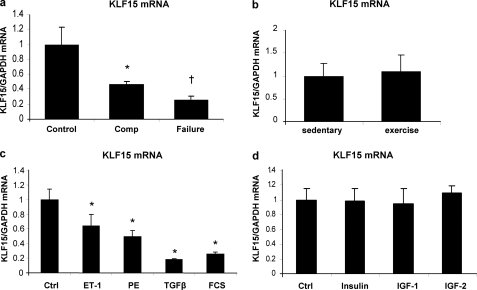
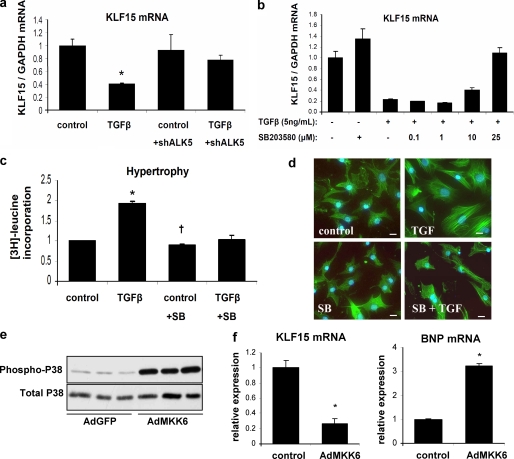
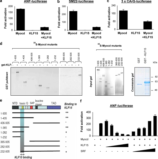
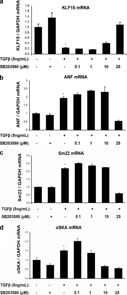
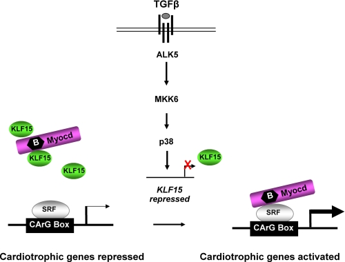
Pathological forms of left ventricular hypertrophy (LVH) often progress to heart failure. Specific transcription factors have been identified that activate the gene program to induce pathological forms of LVH. It is likely that apart from activating transcriptional inducers of LVH, constitutive transcriptional repressors need to be removed during the development of cardiac hypertrophy. Here, we report that the constitutive presence of Krüppel-like factor 15 (KLF15) is lost in pathological hypertrophy and that this loss precedes progression toward heart failure. We show that transforming growth factor-beta-mediated activation of p38 MAPK is necessary and sufficient to decrease KLF15 expression. We further show that KLF15 robustly inhibits myocardin, a potent transcriptional activator. Loss of KLF15 during pathological LVH relieves the inhibitory effects on myocardin and stimulates the expression of serum response factor target genes, such as atrial natriuretic factor. This uncovers a novel mechanism where activated p38 MAPK decreases KLF15, an important constitutive transcriptional repressor whose removal seems a vital step to allow the induction of pathological LVH.


Figures

FIGURE 1.
FIGURE 1.
Loss of KLF15 is specific for pathological hypertrophy and is mediated by the TGFβ1-p38 MAPK pathway. a, KLF15 expression is down-regulated in cardiac biopsies of hypertensive Ren-2 rats. KLF15 is decreased more in the hearts of the rats that after the biopsy progressed to heart failure, as compared with the hypertrophied hearts from the rats that remained compensated after the biopsy (n = 4–6 rats per group; *, p < 0.05 compared with control group; †, p < 0.02 compared with the compensated (Comp) hypertrophy group). b, in hypertrophic myocardium from rats that had undergone exercise training for 10 weeks, 5 days per week (n = 5), KLF15 expression was not altered compared with control rat hearts (n = 7). c, KLF15 mRNA expression is decreased in cultured neonatal rat cardiomyocytes in response to several hypertrophic stimuli. Cardiomyocytes were serum-starved overnight and then stimulated with endothelin 1 (ET-1) (100 nm), phenylephrine (PE) (50 μm), TGFβ (10 ng/ml), and 10% fetal calf serum (FCS) for 24 h. Ctrl, control. KLF15 levels were detected using quantitative real time PCR. (*, p < 0.01 compared with control cells, n = 3 per group). d, KLF15 mRNA levels do not decrease on stimulation of cultured neonatal rat cardiomyocytes with IGF-I (100 nm), IGF-II (100 nm), or insulin (100 nm) for 24 h after 24 h of serum starvation (n = 3 per group).
FIGURE 2.
FIGURE 2.
In vitro activation of p38 MAPK is both necessary and sufficient to decrease KLF15 levels. a, TGFβ regulates KLF15 expression in vitro. Neonatal rat cardiomyocytes were infected with a lentivirus containing shRNA against ALK5 or with a control lentivirus. TGFβ decreased expression of KLF15 after control virus but had no effect in the cells treated with the shRNA. (n = 3/group; *, p < 0.01 compared with control group; †, p < 0.01 compared with TGFβ-treated cells without shRNA against ALK5. b, specific inhibitor of p38 MAPK, SB203580, abolishes the TGFβ-induced down-regulation of KLF15 in cardiomyocytes in a dose-dependent manner. c, inhibition of p38 MAPK prevents TGFβ-induced hypertrophy, measured by [3H]leucine incorporation (n = 3/group; *, p < 0.05 compared with control group; †, p < 0.05 compared with TGFβ-treated cells without SB203580 (SB) treatment), and d, inhibition of p38 MAPK prevents TGFβ-induced increase in cell size as visualized by phalloidin staining of F-actins. Bars in panels represent 50 μm. e, Western blot analysis shows an increase in phosphorylated p38 MAPK after infection of cardiomyocytes with constitutively active adMKK6, the upstream kinase of p38. f, adenoviral overexpression of MKK6 resulted in decreased KLF15 mRNA levels and increased expression of the hypertrophy marker BNP (n = 3/group; *, p < 0.05 compared with control group).
FIGURE 3.
FIGURE 3.
Binding of KLF15 to the basic region of myocardin results in repression of myocardin-responsive reporters. KLF15 inhibits myocardin activity. a–c, myocardin activates the following SRF-dependent reporters: ANF, SM22, and the 3×CArG-luciferase. This activity is blocked by KLF15. COS cells were transfected with the luciferase reporters and expression vectors encoding myocardin-935 and KLF15. Luciferase activity is expressed as fold change over the empty expression vector, pcDNA3.1 (n = 3, mean ± S.D.). d, GST pulldown assays show that KLF15 associates with myocardin. 35S-Labeled myocardin mutant proteins were translated in vitro and incubated with GST-KLF15 fusion protein. Proteins were captured on glutathione-agarose beads and analyzed by SDS-PAGE. The input lanes contain 10% of the amount of 35S-labeled myocardin protein in the pulldown lanes. GST-KLF15 fusion protein is shown on a Coomassie gel on the right. e, schematic representation of full-length myocardin (1–935 amino acids) and myocardin mutants used for GST pulldown (as shown in d) and their binding to KLF15 are indicated to the right. The shaded box represents the region that is necessary for KLF15 binding. NTD, N-terminal domain; Q, glutamine-rich domain; TAD, transactivation domain. f, KLF15 competes with SRF for myocardin interaction. COS cells were transfected with ANF luciferase and expression vectors encoding myocardin-935 and increasing concentrations of KLF15 and SRF as indicated. Luciferase activity expressed as fold change over the empty expression vector, pcDNA3.1, decreases dose dependently with increasing amounts of KLF15. (n = 3, mean ± S.D.)
FIGURE 4.
FIGURE 4.
Induction of myocardin/SRF target genes by TGFβ-p38 MAPK signaling. Quantitative real time PCR demonstrates that SRF targets, such as ANF, SM22, and α-skeletal actin are induced in cardiomyocytes in response to TGFβ treatment (24 h, 10 ng/ml serum-free medium), whereas KLF15 mRNA levels are down-regulated (n = 3/group; *, p < 0.05). This increase in SRF targets by TGFβ is counteracted by the p38 inhibitor SB203580.
FIGURE 5.
FIGURE 5.
Proposed mechanism of action of TGFβ-p38 MAPK-KLF15 cascade to explain induction of cardiomyocyte hypertrophy. KLF15 competes with SRF for a common docking site within myocardin, thereby limiting myocardin to activate cardiotrophic genes in normal adult myocytes. Growth factors repress KLF15 expression, allowing myocardin to bind SRF and activate cardiotrophic genes.

References

    1. Oka T., Xu J., Molkentin J. D. (2007) Semin. Cell Dev. Biol. 18, 117–131 - PMC - PubMed
    1. Oka T., Maillet M., Watt A. J., Schwartz R. J., Aronow B. J., Duncan S. A., Molkentin J. D. (2006) Circ. Res. 98, 837–845 - PubMed
    1. Zhang X., Azhar G., Chai J., Sheridan P., Nagano K., Brown T., Yang J., Khrapko K., Borras A. M., Lawitts J., Misra R. P., Wei J. Y. (2001) Am. J. Physiol. Heart Circ. Physiol. 280, H1782–H1792 - PubMed
    1. Wang D., Chang P. S., Wang Z., Sutherland L., Richardson J. A., Small E., Krieg P. A., Olson E. N. (2001) Cell 105, 851–862 - PubMed
    1. Pipes G. C., Creemers E. E., Olson E. N. (2006) Genes Dev. 20, 1545–1556 - PubMed

Publication types

MeSH terms