. 2010 Aug 13;285(33):25645-53.
doi: 10.1074/jbc.M110.104976. Epub 2010 Jun 21.

Store-operated Ca2+ entry in malignant hyperthermia-susceptible human skeletal muscle

Affiliations

Store-operated Ca2+ entry in malignant hyperthermia-susceptible human skeletal muscle

Adrian M Duke et al. J Biol Chem. .

Abstract

In malignant hyperthermia (MH), mutations in RyR1 underlie direct activation of the channel by volatile anesthetics, leading to muscle contracture and a life-threatening increase in core body temperature. The aim of the present study was to establish whether the associated depletion of sarcoplasmic reticulum (SR) Ca(2+) triggers sarcolemmal Ca(2+) influx via store-operated Ca(2+) entry (SOCE). Samples of vastus medialis muscle were obtained from patients undergoing assessment for MH susceptibility using the in vitro contracture test. Single fibers were mechanically skinned, and confocal microscopy was used to detect changes in [Ca(2+)] either within the resealed t-system ([Ca(2+)](t-sys)) or within the cytosol. In normal fibers, halothane (0.5 mM) failed to initiate SR Ca(2+) release or Ca(2+)(t-sys) depletion. However, in MH-susceptible (MHS) fibers, halothane induced both SR Ca(2+) release and Ca(2+)(t-sys) depletion, consistent with SOCE. In some MHS fibers, halothane-induced SR Ca(2+) release took the form of a propagated wave, which was temporally coupled to a wave of Ca(2+)(t-sys) depletion. SOCE was potently inhibited by "extracellular" application of a STIM1 antibody trapped within the t-system but not when the antibody was denatured by heating. In conclusion, (i) in human MHS muscle, SR Ca(2+) depletion induced by a level of volatile anesthetic within the clinical range is sufficient to induce SOCE, which is tightly coupled to SR Ca(2+) release; (ii) sarcolemmal STIM1 has an important role in regulating SOCE; and (iii) sustained SOCE from an effectively infinite extracellular Ca(2+) pool may contribute to the maintained rise in cytosolic [Ca(2+)] that underlies MH.


Figures
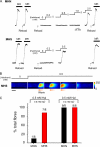
FIGURE 1.
FIGURE 1.
Differential sensitivity of cytosolic SR Ca2+ release to halothane in MHS and MHN fibers. A, typical example showing an endogenously Ca2+-loaded MHN fiber, which was initially exposed to a solution containing 20 mm caffeine and 20 μm Mg2+ to induce a maximal release (MR) of Ca2+ from the SR, resulting in a transient increase in fluo-3 fluorescence within the cytosol (upper panel). The SR was then reloaded to the same by exposure to a solution with a free [Ca2+] of ∼200 nm, and another maximal Ca2+ release was induced. After reloading the SR again, the MHN fiber was subjected to stepwise increases in [halothane] until SR Ca2+ release occurred at 2 mm in this example. When the same protocol was applied to MHS fibers, the threshold for halothane-induced Ca2+ release was consistently ≤0.5 mm (lower panel). B, sequential x-y confocal images (0.83 Hz) from an MHS fiber in which halothane exposure induced a slow propagated Ca2+ wave. Scale bar indicates 80 μm. C, cumulative data showing the proportion of MHS and MHN fibers responding to 0.5 mm halothane in the presence of 0.8 or 0.2 mm Mg2+. The total number of halothane-responsive fibers (one per patient) is indicated above each bar.
FIGURE 2.
FIGURE 2.
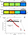
Properties of halothane-induced SOCE. A, typical x-y confocal images of fluo-5N fluorescence obtained with the dye trapped within the resealed t-tubules of a mechanically skinned MHN or MHS fiber. In MHN fibers (upper panel), the introduction of 0.5 mm halothane had no apparent effect on fluo-5N fluorescence within 60 s. However, after initiation of a maximal SR Ca2+ release (MR) by reducing in the free [Mg2+] to 20 μm, t-tubule fluorescence decreased markedly. In MHS fibers (lower panel), the introduction of 0.5 mm halothane resulted in an initial increase in fluo-5N fluorescence followed by a decrease, which approached a new steady state after ∼60 s. Thereafter, initiation of a maximal SR Ca2+ release induced a further decrease in fluo-5N fluorescence. B, cumulative data showing the mean fluorescence obtained from sequential x-y images before and during exposure to halothane or maximal SR Ca2+ release (MR), in MHS (n = 8, red circles) or MHN (n = 8, black circles) human skeletal muscle fibers. Each data point represents mean ± S.E. * indicates significantly different from control, p < 0.05.
FIGURE 3.
FIGURE 3.
Halothane-induced wave of SOCE in human MHS fibers. Sequential surface plots of x-y confocal images taken from an MHS fiber (0.83 Hz), which exhibited a propagated wave of t-tubule [Ca2+] change following exposure to 0.5 mm halothane, are shown. An initial wave front of raised [Ca2+]t-sys was followed by sustained [Ca2+]t-sys depletion. Similar results were obtained in two other preparations.
FIGURE 4.
FIGURE 4.
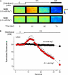
Effect of reduced [ Mg2+] on the response of MHN fibers to halothane. A, typical x-y confocal images of fluo-5N fluorescence within the resealed t-tubules of a skinned MHN fiber in the presence of 0.8 mm (upper panel) or 0.2 mm Mg2+ (lower panel) free [Mg2+]. At 0.8 mm Mg2+, halothane had no apparent effect on fluo-5N fluorescence, whereas a subsequent maximal SR Ca2+ release (MR) induced a sustained decrease in [Ca2+]t-sys. In the presence of 0.2 mm Mg2+, the introduction of halothane resulted in a transient increase in [Ca2+]t-sys followed by a sustained decrease. B, cumulative data showing the mean fluorescence obtained from sequential x-y images before and during exposure to 0.5 mm halothane in the presence of 0.8 mm (n = 8, red circles) or 0.2 mm (n = 8, black circles) free Mg2+. Each data point represents mean ± S.E. * indicates significantly different from control, p < 0.05.
FIGURE 5.
FIGURE 5.
Effects of STIM1 antibody on the response of MHS fibers to halothane. A, x-y confocal images of fluo-5N fluorescence within the resealed t-tubules of a skinned MHS fiber in which a STIM1 antibody had been introduced into the t-system prior to skinning. In this case, exposure to halothane was followed by a characteristic increase in [Ca2+]t-sys (upper panel). However, fluo-5N fluorescence then returned slowly toward the control level, and there was little evidence of a sustained decrease within 60 s or subsequently after a maximal SR Ca2+ release (MR). This procedure was repeated in an MHS fiber using a sample of the antibody, which had been denatured by heating (lower panel). After heat treatment, application of halothane resulted in a typical SOCE response. B, cumulative data showing the mean fluorescence obtained from sequential x-y images in MHS fibers with either the active STIM1 antibody (n = 8, red triangles) or the denatured antibody (n = 8, open squares) trapped within the t-system. * indicates statistically different from control level, p < 0.05. C, representative Western blot from human vastus medialis muscle. STIM1 is indentified in whole muscle human homogenate and is also enriched in the buoyant fraction 6, which contains sarcolemmal lipid rafts and caveolae. β-Adaptin (a marker of non-raft membranes) is present at low levels in fractions 5 and 6, whereas caveolin-3 (a marker of caveolae) is enriched. Fractions 7–12 are mostly non-raft membranes and cytosolic proteins. Unlike some other caveolin isoforms, caveolin-3 is found outside buoyant membrane fractions as observed here in fractions 9–12 (35).

References

    1. Launikonis B. S., Barnes M., Stephenson D. G. (2003) Proc. Natl. Acad. Sci. U.S.A. 100, 2941–2944 - PMC - PubMed
    1. Kurebayashi N., Ogawa Y. (2001) J. Physiol. 533, 185–199 - PMC - PubMed
    1. Launikonis B. S., Ríos E. (2007) J. Physiol. 583, 81–97 - PMC - PubMed
    1. Posterino G. S., Lamb G. D. (2003) J. Physiol. 551, 219–237 - PMC - PubMed
    1. Launikonis B. S., Zhou J., Royer L., Shannon T. R., Brum G., Ríos E. (2006) Proc. Natl. Acad. Sci. U.S.A. 103, 2982–2987 - PMC - PubMed

Publication types