. 2010 Jun 28;189(7):1143-55.
doi: 10.1083/jcb.201001013. Epub 2010 Jun 21.

Dual recognition of CENP-A nucleosomes is required for centromere assembly

Affiliations

Dual recognition of CENP-A nucleosomes is required for centromere assembly

Christopher W Carroll et al. J Cell Biol. .

Abstract

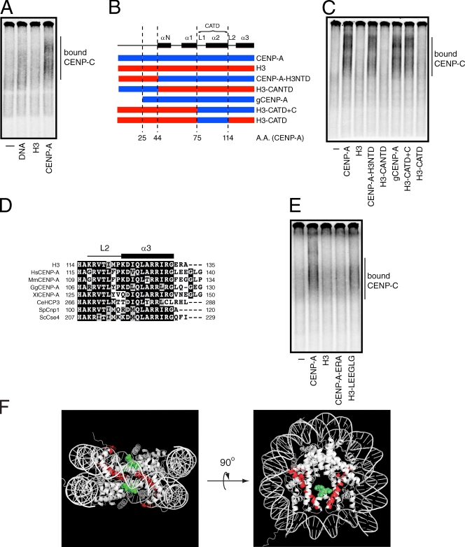
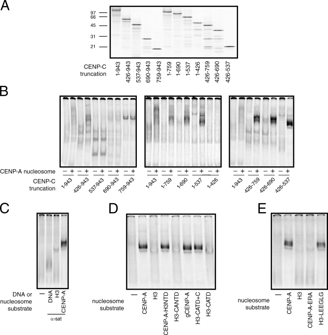
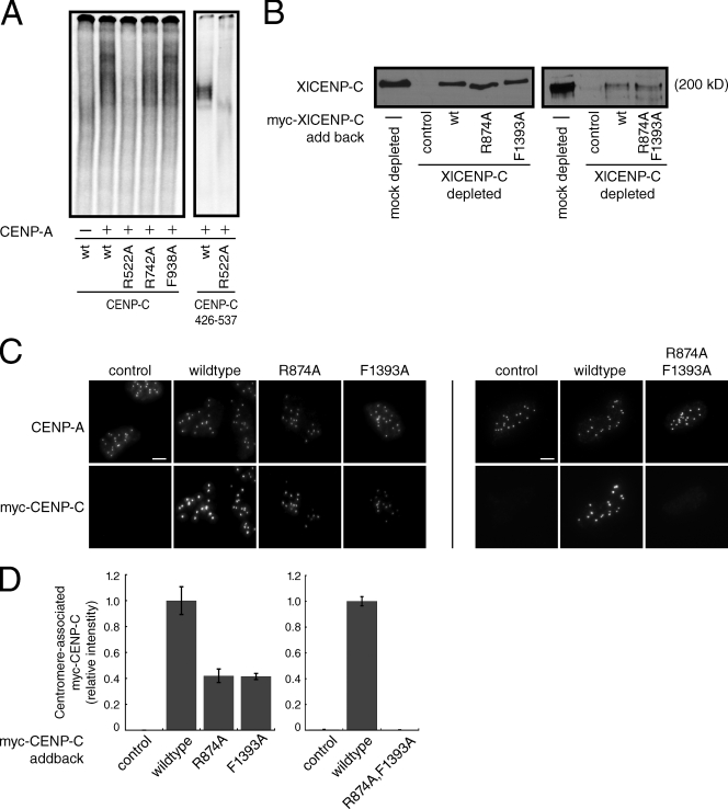
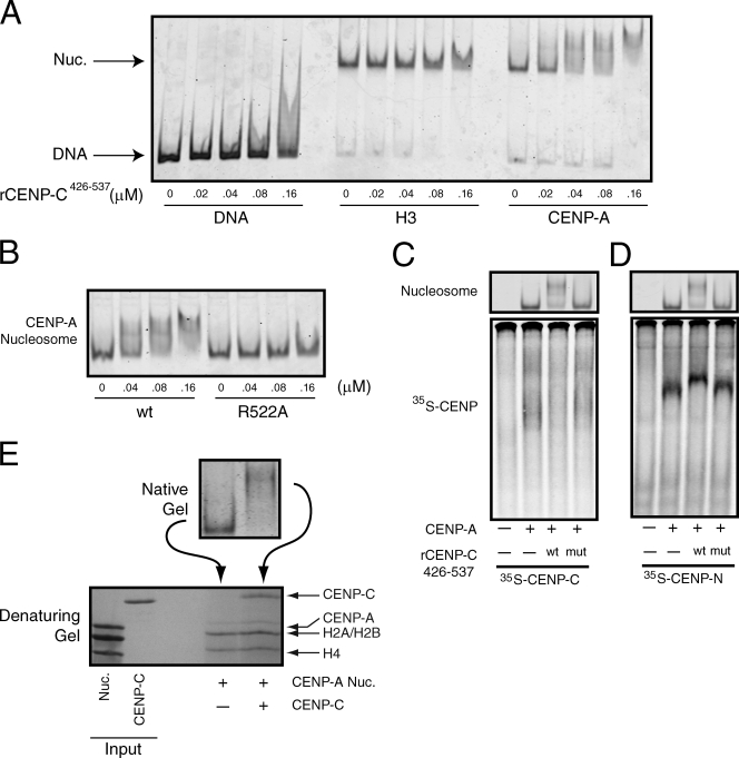
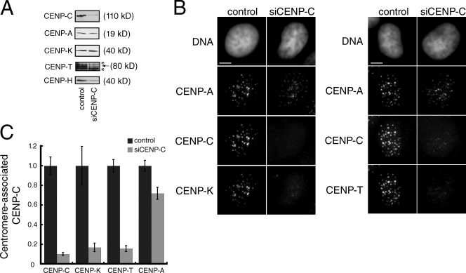
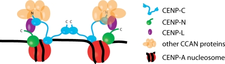
Centromeres contain specialized nucleosomes in which histone H3 is replaced by the histone variant centromere protein A (CENP-A). CENP-A nucleosomes are thought to act as an epigenetic mark that specifies centromere identity. We previously identified CENP-N as a CENP-A nucleosome-specific binding protein. Here, we show that CENP-C also binds directly and specifically to CENP-A nucleosomes. Nucleosome binding by CENP-C required the extreme C terminus of CENP-A and did not compete with CENP-N binding, which suggests that CENP-C and CENP-N recognize distinct structural elements of CENP-A nucleosomes. A mutation that disrupted CENP-C binding to CENP-A nucleosomes in vitro caused defects in CENP-C targeting to centromeres. Moreover, depletion of CENP-C with siRNA resulted in the mislocalization of all other nonhistone CENPs examined, including CENP-K, CENP-H, CENP-I, and CENP-T, and led to a partial reduction in centromeric CENP-A. We propose that CENP-C binds directly to CENP-A chromatin and, together with CENP-N, provides the foundation upon which other centromere and kinetochore proteins are assembled.


Figures

Figure 1.
Figure 1.
CENP-C binds directly to CENP-A nucleosomes. (A) [35S]methionine-labeled CENP-C was incubated alone (−) or in the presence of α-satellite DNA or nucleosomes (300 nM). The mixtures were resolved on a native gel, and [35S]methionine-labeled CENP-C was detected with a phosphorimager. The position of CENP-C bound to DNA or nucleosomes is indicated. (B) A schematic showing the junctions of the CENP-A (blue) and histone H3 (red) chimeras used in this study. The numbers below the diagram refer to amino acids positions (A.A.) within CENP-A. Helical elements (black boxes, labeled α) and loops (labeled L) found within the histone fold of CENP-A and the region encompassing the CATD are shown above for reference. (C) Wild-type nucleosomes or nucleosomes containing CENP-A/H3 chimeras (500 nM) were incubated with [35S]methionine-labeled CENP-C and resolved on a native gel. [35S]methionine-labeled CENP-C alone (−) was used as a negative control. The position of nucleosome-bound CENP-C is indicated. (D) Clustal W alignment of the C terminus of human histone H3 and CENP-A orthologues from humans (Hs), mice (Mm), chickens (Gg), X. laevis (Xl), C. elegans (CeHCP3), S. pombe (SpCnp1), and Saccharomyces cerevisiae (ScCse4). The identical residues within each protein are highlighted with a black background, whereas conservative amino acid changes are colored gray. The first and last amino acid positions of the residues contained within the alignment are shown on the left and right side of each sequence, respectively. The positions of loop 2 (L2) and helix 3 (α3) within CENP-A are shown above for reference. (E) [35S]methionine-labeled CENP-C was incubated alone (−) or in the presence of the indicated nucleosomes and analyzed as in A. (F) The crystal structure of histone H3 is shown. Amino acids positions analogous to the CENP-A CATD (red ribbon) and residues C-terminal to helix 3 (green dots) are highlighted. Only one of the C-terminal extensions is colored green on the right panel for clarity.
Figure 2.
Figure 2.
Identification of a CENP-A recognition module within CENP-C. (A) Full-length (1–943) and various truncation mutants of [35S]methionine-labeled CENP-C were produced in rabbit reticulocyte extracts and resolved on a 12.5% SDS-PAGE gel. The amino acids within CENP-C that are included in each protein are indicated below the gel. The positions of molecular weight markers (kD) are indicated to the left of the gel. (B) Each CENP-C protein was incubated alone (−) or in the presence of 300 nM of CENP-A nucleosomes (+) and resolved on a native gel. (C) CENP-C426–537 was incubated alone (−) or in the presence of α-satellite DNA or the indicated nucleosome (10 nM) and resolved on a native gel. (D and E) CENP-C426–537 was incubated alone (−) or in the presence of the indicated nucleosomes (10 nM) and analyzed as above. The specific regions of CENP-A and H3 included in each chimera are schematized in Fig. 1 B.
Figure 3.
Figure 3.
A conserved arginine residue within CENP-C is required for nucleosome binding and centromere localization. (A) Wild-type (wt) human CENP-C or the indicated point mutant was incubated alone (−) or with CENP-A nucleosomes (+) and resolved on a native gel (left). Alternatively, the wild-type (wt) or R522A CENP-C426–537 fragments were incubated with CENP-A nucleosomes (10 nM). (B) Western blot using an anti–XlCENP-C antibody of mock-depleted or CENP-C–depleted Xenopus extracts containing reticulocyte-produced wild-type XlCENP-C (wt) or the indicated point mutants. Reticulocyte extract lacking myc-CENP-C (−) was used as a control. R874 and F1393 in Xenopus CENP-C are analogous to R522 and F938, respectively, in human CENP-C. The molecular weight of XlCENP-C is indicated. (C) Isolated sperm nuclei from CENP-C–depleted Xenopus extracts containing the indicated myc-CENP-C protein were stained with anti–XlCENP-A and anti-myc antibodies. Images are maximum-intensity projections of z stacks collected at 0.2-µM steps. Bars, 5 µM. (D) Quantification of centromere-associated myc-CENP-C from C. Error bars show the SEM from three independent experiments (>100 centromeres were counted for each condition in each experiment).
Figure 4.
Figure 4.
CENP-C and CENP-N bind to different sites on the same CENP-A nucleosomes. (A) α-Satellite DNA or reconstituted nucleosomes (10 nM) were incubated with the indicated concentration of rCENP-C426–537 and resolved on native gels. DNA or nucleosomes were visualized after staining with SYBR gold. (B) CENP-A nucleosomes (10 nM) were incubated with the indicated concentration of wild-type (wt) or mutant (R522A) rCENP-C426–537 and resolved on a native gel. (C) [35S]methionine-labeled CENP-C was incubated alone (−) or in the presence of 150 nM of CENP-A nucleosomes (+). In addition, binding reactions contained wild-type (wt) or the R522A mutant (mut) rCENP-C426–537 (300 nM), or buffer alone (−). The gel was stained with ethidium bromide to visualize nucleosomes (top) and was subsequently dried and scanned on a phosphorimager to visualize the labeled CENP-C (bottom). (D) An identical experiment to C except that [35S]methionine-labeled CENP-N was used. (E) Isolated nuclei were digested with micrococcal nuclease, and wild-type or R11A mutant GFP–CENP-N was immunoprecipitated using anti-GFP antibodies. Each immunoprecipitate was probed with the indicated antibodies. Control cells don’t express GFP–CENP-N.
Figure 5.
Figure 5.
CENP-C is required for centromere assembly. (A) Western blotting of whole-cell extracts prepared from mock-depleted (control) or CENP-C–depleted HeLa cells. The antibody used for each Western blot is indicated on the left. Background bands (*) were present above and below the CENP-T band, which is indicated with an arrow. The molecular weight of each protein is indicated. (B) Representative images from control or mock-depleted cells stained with Hoechst (DNA) or the indicated antibody. Images are maximum-intensity projections of z stacks collected at 0.2-µM steps. Bars, 5 µM. (C) Quantification of the relative levels at centromeres of the indicated protein in mock- or CENP-C–depleted cells. For each antigen, >20 cells and >200 centromeres were quantified. Error bars indicate the SEM from three independent experiments.
Figure 6.
Figure 6.
A model for centromere assembly in human cells. CENP-A (red), CENP-C (cyan), CENP-N (green), and CENP-L (magenta) nucleosomes are shown. Other CCAN proteins are colored orange. The N and C termini of CENP-C are indicated, as is the C terminus of CENP-N.

References

    1. Amano M., Suzuki A., Hori T., Backer C., Okawa K., Cheeseman I.M., Fukagawa T. 2009. The CENP-S complex is essential for the stable assembly of outer kinetochore structure. J. Cell Biol. 186:173–182 10.1083/jcb.200903100 - DOI - PMC - PubMed
    1. Ando S., Yang H., Nozaki N., Okazaki T., Yoda K. 2002. CENP-A, -B, and -C chromatin complex that contains the I-type alpha-satellite array constitutes the prekinetochore in HeLa cells. Mol. Cell. Biol. 22:2229–2241 10.1128/MCB.22.7.2229-2241.2002 - DOI - PMC - PubMed
    1. Black B.E., Foltz D.R., Chakravarthy S., Luger K., Woods V.L., Jr., Cleveland D.W. 2004. Structural determinants for generating centromeric chromatin. Nature. 430:578–582 10.1038/nature02766 - DOI - PubMed
    1. Black B.E., Jansen L.E., Maddox P.S., Foltz D.R., Desai A.B., Shah J.V., Cleveland D.W. 2007. Centromere identity maintained by nucleosomes assembled with histone H3 containing the CENP-A targeting domain. Mol. Cell. 25:309–322 10.1016/j.molcel.2006.12.018 - DOI - PubMed
    1. Blower M.D., Karpen G.H. 2001. The role of Drosophila CID in kinetochore formation, cell-cycle progression and heterochromatin interactions. Nat. Cell Biol. 3:730–739 10.1038/35087045 - DOI - PMC - PubMed

Publication types