. 2010 Aug;30(16):4060-76.
doi: 10.1128/MCB.01399-09. Epub 2010 Jun 21.

In vitro reconstitution of PHO5 promoter chromatin remodeling points to a role for activator-nucleosome competition in vivo

Affiliations

In vitro reconstitution of PHO5 promoter chromatin remodeling points to a role for activator-nucleosome competition in vivo

Franziska Ertel et al. Mol Cell Biol. 2010 Aug.

Abstract

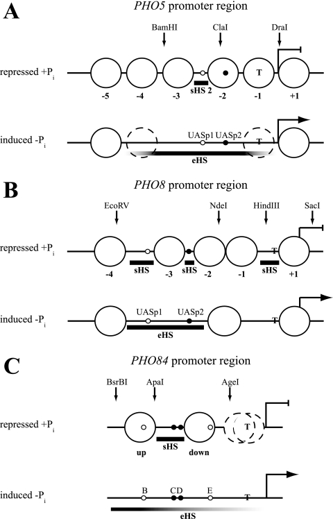
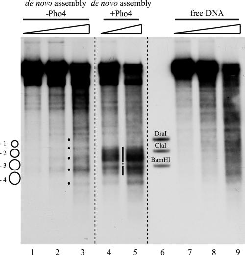
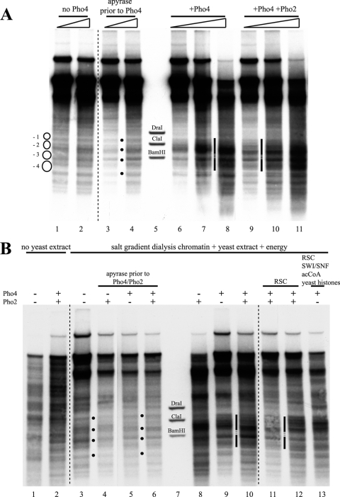
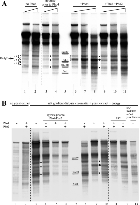
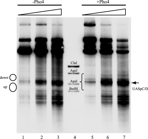
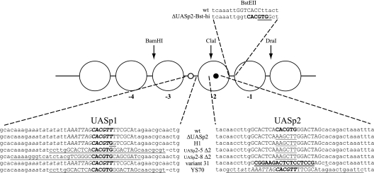
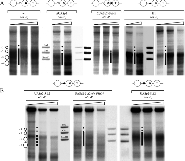
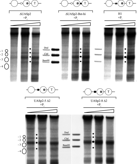
The yeast PHO5 promoter is a classical model for studying the role of chromatin in gene regulation. To enable biochemical dissection of the mechanism leading to PHO5 activation, we reconstituted the process in vitro. Positioned nucleosomes corresponding to the repressed PHO5 promoter state were assembled using a yeast extract-based in vitro system. Addition of the transactivator Pho4 yielded an extensive DNase I-hypersensitive site resembling induced PHO5 promoter chromatin. Importantly, this remodeling was energy dependent. In contrast, little or no chromatin remodeling was detected at the PHO8 or PHO84 promoter in this in vitro system. Only the PHO5 promoter harbors a high-affinity intranucleosomal Pho4 binding site (UASp) where Pho4 binding can compete with nucleosome formation, prompting us to test the importance of such competition for chromatin remodeling by analysis of UASp mutants in vivo. Indeed, the intranucleosomal location of the UASp element was critical, but not essential, for complete remodeling at the PHO5 promoter in vivo. Further, binding of just the Gal4 DNA binding domain to an intranucleosomal site could increase PHO5 promoter opening. These data establish an auxiliary role for DNA binding competition between Pho4 and histones in PHO5 promoter chromatin remodeling in vivo.


Figures

FIG. 1.
FIG. 1.
Schematics of the nucleosomal organization at the PHO5, PHO8, and PHO84 promoter regions in the repressed (+Pi) and induced (−Pi) states. Large circles denote positioned nucleosomes, which are numbered relative to the ATG at the PHO5 promoter and relative to the short hypersensitive region just upstream of the ATG at the PHO8 promoter. The positioned nucleosomes flanking the short hypersensitive region at the PHO84 promoter are labeled “up” and “down” according to reference . Stippled circles stand for nucleosomes that are partially remodeled (PHO5, −Pi) (2, 10, 11, 34) or show ambiguous positioning (PHO84, +Pi) (67). Bold horizontal bars demarcate short (sHS) or extensive (eHS) DNase I-hypersensitive sites. Fading of the bar symbolizes less hypersensitivity. The positions of high-affinity Pho4 binding sites (UASp2 at PHO5 and PHO8, C and D at PHO84) are indicated by small filled circles, and the positions of low-affinity sites (UASp1 at PHO5 and PHO8, B and E at PHO84) are indicated by small open circles. The positions of the TATA box (T), the ATG (broken blunt and broken pointed arrows for repressed and induced states, respectively), and the restriction sites used for generating marker fragments are shown. The schematics are mainly based on previous publications on PHO5 (2, 29), PHO8 (3), and PHO84 (67). For a comparison of these schematics with data from other sources, see reference .
FIG. 2.
FIG. 2.
Pho4-induced generation of a hypersensitive site at the PHO5 promoter during de novo assembly in vitro. DNase I indirect end-labeling analysis of the PHO5 promoter region in chromatin assembled de novo with yeast extract (from pho4 strain YS22) in vitro with or without the addition of exogenous Pho4 and in free DNA. The schematic on the left denotes positioned nucleosomes as in the schematic of the PHO5 promoter in Fig. 1A. Black dots between lanes 2 and 3 mark the bands corresponding to the linker regions between the positioned nucleosomes. The vertical bar between lanes 4 and 5 highlights the hypersensitive region generated by the addition of Pho4. The stippled region interrupting the vertical bar points to a region of maintained protection from DNase I. Ramps above the lanes stand for increasing DNase I concentrations. Marker fragments were generated by double digests of DraI, ClaI, and BamHI, each with ApaI. All samples were electrophoresed in lanes of the same gel. Stippled lines between lanes show where lanes were moved next to each other using Adobe Photoshop CS2.
FIG. 3.
FIG. 3.
Pho4- and energy-dependent remodeling of prepositioned nucleosomes at the PHO5 promoter in vitro. (A) DNase I indirect end-labeling analysis of the PHO5 promoter region in chromatin treated as indicated after preassembly by salt gradient dialysis and incubation with yeast extract (from strain YS27 cpf1) in the presence of energy. The schematic, black dots, vertical bars, stippled lines, ramps, and markers are as in Fig. 2. (B) Same as panel A but with treatment of salt gradient dialysis chromatin as indicated above the lanes. Only for the sample in lane 12 were Pho4 and Pho2 added at the same time as the yeast extract. For all of the samples in lanes 3 to 6, 8 to 11, and 13, salt gradient dialysis chromatin was first treated with yeast extract and energy to yield the pattern shown in lane 3 and then further treated for 1 h at 30°C as indicated. In all DNase I mapping experiments, a range of DNase I concentrations was used, but due to space limitations, only one representative concentration is shown for each condition. acCoA, acetyl-CoA.
FIG. 4.
FIG. 4.
Pho4-induced remodeling at the PHO5 promoter in vitro is similar to but less extensive than in vivo. (A) Same as Fig. 3A but with in vivo samples from wild-type strain CY337 corresponding to the repressed (+Pi) and induced (−Pi) states of the PHO5 promoter electrophoresed alongside in vitro samples. Note that the vertical bar between lanes 6 and 7 highlighting the in vitro hypersensitive region is interrupted by a short stippled stretch at a position of DNA protection that is fully accessible in the induced state in vivo (bar between lanes 8 and 9). (B) ClaI accessibility values for in vitro-assembled chromatin treated as indicated. For columns 4 to 12, Pho4 and/or Pho2 were added after the salt gradient dialysis chromatin was incubated with yeast extract. For columns 13 to 16, they were added together with the yeast extract. Error bars show the variation of two or three independent experiments starting from the same salt gradient dialysis chromatin preparation. In the case of three experiments, the error bars correspond to the standard deviation.
FIG. 5.
FIG. 5.
Little or no Pho4-induced chromatin remodeling at the PHO8 promoter in vitro. Panel A shows the same blot as in Fig. 3A, and panel B shows the same blot as in Fig. 3B but with both rehybridized for the PHO8 promoter region. The schematic on the left in panel A corresponds to Fig. 1B. The brace between lanes 5 and 6 shows the region that becomes hypersensitive upon induction in vivo (3). Marker fragments were generated by double digests of EcoRV, NdeI, HindIII, and SacI (panel A) or EcoRV, HindIII, and XhoI (panel B), each with BglII. Dots between lanes mark the bands characteristic for the PHO8 promoter chromatin pattern of the repressed state. Larger dots mark increased sensitivity of the band corresponding to the UASp2 Pho4 binding site.
FIG. 6.
FIG. 6.
Little or no Pho4-induced chromatin remodeling at the PHO84 promoter in vitro. DNase I indirect end-labeling analysis of the PHO84 promoter region in chromatin preassembled by salt gradient dialysis, incubated with yeast extract (from strain YS27 cpf1) in the presence of energy and then left untreated or treated with Pho4, as indicated. The schematic on the left corresponds to positioned nucleosomes, as in Fig. 1C. The brace between lanes 4 and 5 shows the region that becomes hypersensitive upon induction in vivo (67). Marker fragments were generated by double digests of ClaI, AgeI, ApaI, and BsrBI, each with SspI. Ramps above the lanes denote increasing DNase I concentrations.
FIG. 7.
FIG. 7.
PHO5 promoter UASp mutants. Schematic of PHO5 promoter mutants with altered UASp elements. The bases of the core hexanucleotide E box of the UASp elements are shown in bold uppercase letters. The UASp2 E box was deleted by conversion to a HindIII site. Mutated DNA regions are underlined. Uppercase bases at the endogenous UASp1 and UASp2 positions correspond to the Pho4 dimethyl sulfate footprint region, and italic bases correspond to the Pho2 dimethyl sulfate footprint region (3, 6, 65). All underlined bases at the UASp1 position of the UASp2-8 Δ2 mutant stem from the UASp2 region of the PHO8 promoter. The bold underlined uppercase bases at the UASp2 position in variant 31 correspond to the introduced Gal4 binding site. The A-to-T point mutation downstream introduced a SacI site. The BstEII site in the wild-type sequence of the linker between the −1 and −2 nucleosomes is indicated in uppercase letters.
FIG. 8.
FIG. 8.
The intranucleosomal location of a UASp element in the −2 nucleosome is important but not essential for PHO5 promoter chromatin opening in vivo. DNase I indirect end-labeling analysis of the PHO5 promoter region in the (A) wild type (wt) (CY337), ΔUASp2 (CY341), ΔUASp2-Bst-hi (CY337 FE1600), and H1 (CY337 EB1615) (35) configurations and in the (B) UASp2-5 Δ2 (CY339 ura3 pCB-UASp2-5 Δ2), and UASp2-8 Δ2 (CY339 ura3 pCB-UASp2-8 Δ2) configurations after overnight (o/n) incubation in phosphate-free (−Pi) medium. For the UASp2-5 Δ2 mutant, an experiment with incubation in phosphate-free medium for 40 h, i.e., two times overnight, and one experiment with overexpression (o/x) of PHO4 are shown. Small dots between lanes mark the bands corresponding to the linker regions flanking the positioned −1 nucleosome as shown in the schematic on the left, and vertical bars between the lanes highlight the extent of a hypersensitive region, stippled if less extensive. For UASp2-5 Δ2, the stippled vertical line denotes less-pronounced hypersensitivity in the region of the −3 and −4 nucleosomes and the large dot marks the increased hypersensitivity of the linker between the −2 and −3 nucleosomes. Samples from the same chromatin preparation but from two different gels are shown for the H1 mutant promoter. Schematics above the panels are as in Table 1. Markers, ramps, and schematics next to the gels are as in Fig. 2.
FIG. 9.
FIG. 9.
PHO5 promoter UASp mutants show mostly undisturbed chromatin patterns in the repressed state. DNase I indirect end-labeling analysis of the indicated PHO5 promoter mutant regions under repressive (+Pi) conditions. The strains are the same as in Fig. 8. The schematics above the lanes are the same as in Table 1. The schematics on the left and the marker bands are the same as in Fig. 2. Small dots between the lanes mark the linker regions between positioned nucleosomes. Larger dots mark enhanced hypersensitivity of sHS2 in mutants compared to the wild type. Ramps above the lanes denote increasing DNase I concentrations.

Similar articles

Cited by

References

    1. Adkins, M. W., S. R. Howar, and J. K. Tyler. 2004. Chromatin disassembly mediated by the histone chaperone Asf1 is essential for transcriptional activation of the yeast PHO5 and PHO8 genes. Mol. Cell 14:657-666. - PubMed
    1. Almer, A., H. Rudolph, A. Hinnen, and W. Hörz. 1986. Removal of positioned nucleosomes from the yeast PHO5 promoter upon PHO5 induction releases additional upstream activating DNA elements. EMBO J. 5:2689-2696. - PMC - PubMed
    1. Barbarić, S., K. D. Fascher, and W. Hörz. 1992. Activation of the weakly regulated PHO8 promoter in S. cerevisiae: chromatin transition and binding sites for the positive regulator protein Pho4. Nucleic Acids Res. 20:1031-1038. - PMC - PubMed
    1. Barbarić, S., T. Luckenbach, A. Schmid, D. Blaschke, W. Hörz, and P. Korber. 2007. Redundancy of chromatin remodeling pathways for the induction of the yeast PHO5 promoter in vivo. J. Biol. Chem. 282:27610-27621. - PubMed
    1. Barbarić, S., M. Münsterkötter, C. Goding, and W. Hörz. 1998. Cooperative Pho2-Pho4 interactions at the PHO5 promoter are critical for binding of Pho4 to UASp1 and for efficient transactivation by Pho4 at UASp2. Mol. Cell. Biol. 18:2629-2639. - PMC - PubMed

Publication types

MeSH terms

LinkOut - more resources