. 2010 Aug;177(2):608-21.
doi: 10.2353/ajpath.2010.091122. Epub 2010 Jun 21.

Inhibition of NF-kappaB signaling reduces virus load and gammaherpesvirus-induced pulmonary fibrosis

Affiliations

Inhibition of NF-kappaB signaling reduces virus load and gammaherpesvirus-induced pulmonary fibrosis

Laurie T Krug et al. Am J Pathol. 2010 Aug.

Abstract

Idiopathic pulmonary fibrosis (IPF) is a chronic progressive lung disorder of unknown etiology. Several studies have demonstrated an association between pulmonary infection with a herpesvirus and IPF. Based on those observations, we have developed a mouse model in which interferon (IFN)gammaR(-/-) mice infected intranasally with murine gammaherpesvirus 68 (MHV68) develop lung fibrosis. We hypothesize that viral load was a critical factor for the development of fibrosis. Because nuclear factor (NF)-kappaB signaling is required to efficiently establish gammaherpesvirus, latency we infected IFNgammaR(-/-) mice with a MHV68 virus that expresses a mutant dominant inhibitor of the NF-kappaB signaling pathway, called IkappaBalphaM. Striking differences were observed at the onset of the chronic infection, which correlated with a decreased virus load in mice infected with MHV68-IkappaBalphaM compared with mice infected with control MHV68 (MHV68-MR). IFNgammaR(-/-) mice infected with MHV68-IkappaBalphaM lacked vasculitis and fibrosis 15 to 120 days post infection. Inhibition of NF-kappaB in MHV68-infected cells of the lungs diminished the expression of the fibrocyte recruiting chemokines monocyte chemoattractant protein 1 (MCP-1) and CXCL12, ameliorated macrophage expression of markers of alternative activation, and failed to increase expression of the integrin alphavbeta6, which is implicated in the activation of the profibrotic factor TGF-beta. Thus, the inhibition of NF-kappaB signaling in the infected lung cells of IFNgammaR(-/-) mice reduces virus persistence and ameliorates profibrotic events. Host determinants of latency might therefore represent new therapeutic targets for gammaherpesvirus-associated pulmonary fibrosis.


Figures

Figure 1
Figure 1
NF-κB activation in response to MHV68 infection can be suppressed by MHV68-IκBαM in vivo. A: Nuclear extracts were prepared from LA-4 cells, murine airway epithelial cells, treated 15 minutes with TNF-α, or infected at an MOI of 5.0 for 24 hours and analyzed by electrophoretic mobility shift assay (EMSA). NF-κB subunits present in the nucleoprotein complexes were identified by supershift analysis using antibodies against the indicated NF-κB subunits. Lines point to specific complexes; bracket indicates supershifted complexes. B: Immunofluorescence of MLE15 cells infected at an MOI of 0.01 48 hpi demonstrates nuclear localization of NF-κB p65 subunit in infected but not uninfected cells. C: Nuclear extracts were prepared from the lungs of a naïve IFNγR−/− mouse (N) or infected (1 and 2) IFNγR−/− mice 7 dpi and analyzed by EMSA. The composition of the complexes was determined by the addition of antibodies against the indicated NF-κB subunits. D: Lung lysates were prepared from NF-κB luc reporter mice mock-infected or infected with MHV68-MR or MHV68-IκBαM at the indicated dpi, normalized by protein content, and subjected to luciferase assay. The data shown represent four to six mice per experimental group. RLU, relative luciferase units. *P < 0.05.
Figure 2
Figure 2
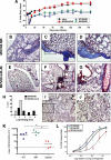
Defective establishment of latency by inhibition of NF-κB in gammaherpesvirus infection protects IFN-γR–deficient mice from virus-induced fibrosis. A: Data are represented as the percentage of the initial body weight (dotted line). More severe illness was observed in MHV68-wild-type and MHV68-MR–infected mice. Data were compiled from two independent experiments (n = 4–18 mice per data point). B and C: Masson trichrome staining of lung slides from two different mice infected with MHV68-MR (marker rescue) at day 120 post infection. Collagen deposition is demonstrated by blue staining. Notice the thickening of the pleura and alveolar walls. Inset in C is shown at higher power view in D. E and F: Masson trichrome staining of lung slides from two different mice infected with MHV68-IκBαM at day 120 post infection. Lymphocytic infiltrates are observed in perivascular areas, but there is no collagen deposition in pleura or alveolar walls. Inset in F is shown at higher power view in G. H: Semiquantitative morphometric analysis of lung histopathology in virus-infected mice with MHV68-MR and MHV68-IκBαM at day 120 of infection. MHV68-MR–infected mice had higher pathology scores corresponding to thickening of the intraalveolar septa and the pleura. In contrast, mice infected with MHV68-IκBαM had lymphocytic infiltrates (n = 13 for MHV68-MR and n = 9 for MHV68-IκBαM). I: Masson trichrome staining of naïve IFNγR−/− mouse. Inset in I is shown at higher power view in J. K: Viral load in the lung was quantified by real-time PCR detection of the virus-genome specific orf50-DNA as compared with cellular GAPDH DNA in 200 ng total DNA prepared from lung tissue of four individual mice infected with MHV68-wild-type (closed squares), MHV68-MR (inverted triangles), and MHV68-IκBαM (open triangles) at 198 dpi. Bars represent geometric mean (*P < 0.01). Dotted line represents detection limit of the assay. L: Persistence in the lungs was analyzed by limiting dilution analysis of disrupted lung tissue isolated from IFNγR−/− mice infected with the indicated viruses 198 dpi. Symbols for each sample represent the mean percentage of 12 wells, positive for cytopathic effect ± the SEM on plating six fourfold dilutions of mechanically disrupted lung tissue from an individual mouse on an indicator MEF monolayer (n = 3–4 mice). Curve fit lines were derived from nonlinear regression analysis. Magnification, ×4 (B, C, E, F, and I).
Figure 3
Figure 3
Inhibition of NF-κB in MHV68 infection does not impair virus replication or inflammation during the acute period of infection in IFNγR−/− mice. A: Acute replication in the lungs of IFNγR−/− mice. On the indicated days post infection, lungs were harvested, disrupted, and titered on NIH 3T12 cells. The data were compiled from one or two experiments with five to ten mice analyzed per experiment. Data are shown as log10 titer, and the bar indicates the geometric mean titer. The dashed line indicates the limit of detection of this assay as log101.7 or 50 pfu/ml of sample homogenate. B: Multistep growth curve in RLE-6TN cells infected with an MOI of 0.01 pfu per cell. Samples were harvested at the indicated time points and titers were determined on NIH3T12 cells. C–F: H&E staining of lung sections from mice infected with MHV68-MR, at 4, 9, and 12 dpi, respectively. Severe inflammation is noted in subpleural areas, around small and medium blood vessels, and surrounding airways by 12 dpi. Inset in E is shown at higher power view in F. G–J: H&E staining of lung sections from mice infected with MHV68- IκBαM, at 4, 9, and 12 dpi, respectively. Inset in I is shown at higher magnification in J. Magnification, ×10 (C–E, G–I).
Figure 4
Figure 4
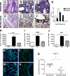
NF-κB inhibition reduces viral load and collagen deposition in the lung tissue of IFNγR−/− mice at the transition from acute to chronic infection. A: H&E staining of lung section from IFNγR−/− mouse infected with MHV68-MR at day 15 pi. B: Masson trichrome staining for collagen deposition of lung section from IFNγR−/− mouse infected with MHV68-MR at day 15 pi. Inset in B is shown in C. D: H&E staining of lung section from IFNγR−/− mouse infected with MHV68-IκBαM at day 15 pi. E: Masson trichrome staining for collagen deposition of lung section from IFNγR−/− mouse infected with MHV68-IκBαM at day 15 pi. Inset in E is shown in F. Notice the increased degree of pleural and alveolar thickening in mice infected with MHV68-MR, as compared with MHV68-IκBαM. G: Semiquantitative morphometric analysis of lung histopathology in virus-infected mice with MHV68-MR and MHV68-IκBαM at 15 dpi. MHV68-MR–infected mice had higher pathology scores corresponding to thickening of the intraalveolar septa. H–J: Quantitative RT-PCR analysis for plasminogen activator 1 (PAI 1), collagen type I (Col1), and smooth muscle actin (α-SMA) transcripts in the lungs of IFNγR−/− mice infected with the indicated viruses at 15 dpi (*P < 0.05, n = 4). K and L: Viral antigen in immunostained frozen lung sections from MHV68-MR versus MHV68-IκBαM–infected IFNγR−/− mice. Insets in K and L are shown in M and N, respectively. O: Quantitative PCR analysis of virus load in the lungs of individual IFNγR−/− mice infected with MHV68-MR (inverted triangles) or MHV68-IκBαM (open triangles) at 15 dpi. Bars represent geometric mean (*P < 0.01). Magnification, ×10 (A–D); ×20 (K and L).
Figure 5
Figure 5
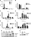
Alternatively activated macrophages as well as chemokine MCP-1 and CXCL12 expression levels are diminished on the infection of IFNγR−/− mice with MHV68-IκBαM. A: Differential cell counts in the bronchoalveolar lavage (BAL) of infected mice at 15 dpi. M indicates mononuclear cells; L, lymphocytes; N, neutrophils; E, eosinophils (*P < 0.01, n = 8). B: Flow cytometry was used to analyzed the CD8+ T cell profile of IFNγR−/− mice uninfected or infected with MHV68-MR or MHV68-IκBαM at 15 dpi. Bars represent the mean percentage of effector (CD44hi, CD62lo) cells; the mean percentage of activated, MHV68-specific (CD44lo, MHV68–p79-peptide specific (tetramer+)) cells; the mean percentage of CD44hi, Vβ4+ populations in gated CD8+ T cells ± SD (n = 3–4 mice per group) at 15 dpi. C–E: TNF-α, IFN-γ, and MCP-1 levels were measured in bronchoalveolar lavage (BAL) fluid from uninfected and MHV68-MR and IκBαM-infected IFNγR−/− mice at the indicated time points post infection. Bars represent mean (n = 5 per group) ± SEM (*P < 0.05). F: Levels of CXCL12 in lung lysates of IFNγR−/− mice after infection with MHV68 determined by ELISA in whole lung lysate. Values represent mean ± SEM (n = 3–8, *P < 0.05). G: Immunoblot analyses for markers of alternative activated macrophages, Arginase 1 and Ym1/2, in lung lysates of naïve mice (N) and mice infected with MHV68-MR and IκBαM at the indicated time points post infection. Each lane represents a different mouse. Blots were stripped and reprobed with an anti–β-actin antibody as loading control. H: Immunoblot assay for Arginase 1 in whole cell extracts from alveolar macrophages isolated from infected MHV68-MR and MHV68-IκBαM mice at day 10 pi.
Figure 6
Figure 6
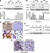
Defective expression and activation of TGF-β in mice infected with MHV68-IκBαM. A: Western blot assay for latent form of TGF-β in BAL collected from naïve (N) and MHV68-MR and MHV68-IκBαM–infected mice at 4 and 9 dpi. Each lane represents a different mouse. Blot was stripped and reprobed with anti-surfactant A (SP-A) antibody to normalize expression of latent TGF-β. B: Immunoblot analysis for the latent and active forms of TGF-β in BAL collected at 15 and 90 dpi. Each lane represents a different mouse. Blot was stripped and reprobed with an anti-surfactant A antibody to normalize expression of latent (white bars) and active (gray bars) TGF-β. Decreased levels of latent and active TGF-β were found in MHV68-IκBαM–infected mice. C: Immunohistochemical analysis of lungs from MHV68-MR and MHV68-IkBαM–infected mice at 90 dpi, using anti–TGF-β specific antibody. Magnification, ×10 (upper); ×100 (lower). Strong positive staining was found in alveolar hyperplasic lung epithelial cells in MHV68-MR–infected mice. D: Immunoblot assay of lung lysates from MHV68-MR and MHV68-IκΒαM–infected mice at the indicated time point post infection using an anti-integrin β6 antibody. Increased levels of integrin β6 were found in samples from MHV68-MR–infected mice. Blot was stripped and reprobed with an anti–β-actin antibody as a loading control. E: Immunostaining for integrin β6 on lung sections from MHV68-MR and -IκΒαM at 15 dpi. High expression for integrin β6 was found in lung sections from MHV68-MR–infected mice. Magnification, ×40.
Figure 7
Figure 7
Defective latency by NF-κB inhibition blunts profibrotic events in MHV68-induced fibrosis. A: MMP9 expression in lung homogenates from naïve and MHV68-MR and MHV68–IκBαM–infected mice at 90 dpi. Each lane represents a different mouse. B: Gelatin zymography of BAL fluid samples from naïve or infected mice at 15 and 90 dpi. Each lane represents a different mouse. Recombinant MMP-9 and MMP-2 were used as controls. High gelatinolytic activity was observed in samples obtained from MHV68-MR–infected mice. C: Quantitative RT-PCR analysis for smooth muscle actin (α-SMA) (*P < 0.05, n = 4). D: Collagen type 1 (Col1) transcripts in the lungs of IFNγR−/− mice infected with the indicated viruses at 90 dpi. E: Immunoblot analysis of collagen 1 in lung lysates of mice infected with MHV68-MR and MHV68-IκBαM at 90 pi. Each lane represents a different mouse. Blot was stripped and reprobed with an anti–β-actin antibody as loading control.

Similar articles

Cited by

References

    1. American Thoracic Society Idiopathic pulmonary fibrosis: diagnosis and treatment. International consensus statement. American Thoracic Society (ATS), and the European Respiratory Society (ERS). Am J Respir Crit Care Med. 2000;161:646–664. - PubMed
    1. Stewart JP, Egan JJ, Ross AJ, Kelly BG, Lok SS, Hasleton PS, Woodcock AA. The detection of Epstein-Barr virus DNA in lung tissue from patients with idiopathic pulmonary fibrosis. Am J Respir Crit Care Med. 1999;159:1336–1341. - PubMed
    1. Egan JJ, Stewart JP, Hasleton PS, Arrand JR, Carroll KB, Woodcock AA. Epstein-Barr virus replication within pulmonary epithelial cells in cryptogenic fibrosing alveolitis. Thorax. 1995;50:1234–1239. - PMC - PubMed
    1. Kelly BG, Lok SS, Hasleton PS, Egan JJ, Stewart JP. A rearranged form of Epstein-Barr virus DNA is associated with idiopathic pulmonary fibrosis. Am J Respir Crit Care Med. 2002;166:510–513. - PubMed
    1. Tang YW, Johnson JE, Browning PJ, Cruz-Gervis RA, Davis A, Graham BS, Brigham KL, Oates JA, Jr, Loyd JE, Stecenko AA. Herpesvirus DNA is consistently detected in lungs of patients with idiopathic pulmonary fibrosis. J Clin Microbiol. 2003;41:2633–2640. - PMC - PubMed

Publication types

MeSH terms