Utilization guidelines for reducing radiation exposure in the evaluation of aneurysmal subarachnoid hemorrhage: A practice quality improvement project
- PMID: 20566813
- PMCID: PMC3130505
- DOI: 10.2214/AJR.09.3786
Utilization guidelines for reducing radiation exposure in the evaluation of aneurysmal subarachnoid hemorrhage: A practice quality improvement project
Abstract
Objective: The purpose of this study was to reduce the cumulative radiation exposure from CT of patients with aneurysmal subarachnoid hemorrhage.
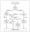
Materials and methods: Baseline data on 30 patients with aneurysmal subarachnoid hemorrhage were collected retrospectively for all CT examinations of the head performed throughout the hospital course. Radiation exposure estimates were obtained by recording dose-length products for each examination. As a departmental practice quality improvement project, an imaging protocol was implemented that included utilization guidelines to reduce radiation exposure in CTA and CT perfusion examinations performed to detect vasospasm in patients with aneurysmal subarachnoid hemorrhage. Ten months after implementation of this protocol, data on 30 additional patients were analyzed. Means, medians, and SD estimates were compared for cumulative radiation exposure and absolute numbers of each examination.
Results: Sixty patients were included in the study: 30 patients at baseline and 30 patients after implementation of the quality improvement plan. These patients underwent 435 CT examinations: 248 examinations at baseline and 187 examinations with the new protocol. With the new algorithm, the mean number of CT examinations per patient was 5.8 compared with 7.8 at baseline, representing a decrease of 25.6%. The number of CT perfusion examinations per patient decreased 32.1%. Overall, there was a 12.1% decrease in cumulative radiation exposure (p > 0.05).
Conclusion: With the structured imaging algorithm, the cumulative radiation exposure and number of CT examinations of the head decreased among patients with aneurysmal subarachnoid hemorrhage because utilization guidelines defined the appropriate imaging time points for detection of vasospasm. Application of these methods to other patient populations with high use of CT may reduce cumulative radiation exposure while the clinical benefits of imaging are maintained.
Figures
Similar articles
-
Radiation exposure in patients with subarachnoid hemorrhage: a quality improvement target.J Neurosurg. 2013 Jul;119(1):215-20. doi: 10.3171/2013.3.JNS12253. Epub 2013 Apr 26. J Neurosurg. 2013. PMID: 23621604
-
Cumulative radiation dose during hospitalization for aneurysmal subarachnoid hemorrhage.AJNR Am J Neuroradiol. 2010 Sep;31(8):1377-82. doi: 10.3174/ajnr.A2132. Epub 2010 May 27. AJNR Am J Neuroradiol. 2010. PMID: 20507932 Free PMC article.
-
Diagnostic Accuracy of Simulated Low-Dose Perfusion CT to Detect Cerebral Perfusion Impairment after Aneurysmal Subarachnoid Hemorrhage: A Retrospective Analysis.Radiology. 2018 May;287(2):643-650. doi: 10.1148/radiol.2017162707. Epub 2018 Jan 8. Radiology. 2018. PMID: 29309735
-
Diagnosing Vasospasm After Subarachnoid Hemorrhage: CTA and CTP.Can J Neurol Sci. 2014 May;41(3):314-9. doi: 10.1017/s031716710001725x. Can J Neurol Sci. 2014. PMID: 24718816 Review.
-
Nonaneurysmal "Pseudo-Subarachnoid Hemorrhage" Computed Tomography Patterns: Challenges in an Acute Decision-Making Heuristics.J Stroke Cerebrovasc Dis. 2018 Sep;27(9):2319-2326. doi: 10.1016/j.jstrokecerebrovasdis.2018.04.016. Epub 2018 Jun 5. J Stroke Cerebrovasc Dis. 2018. PMID: 29884521 Review.
Cited by
-
The why, who, how, and what of communicating CT radiation risks to patients and healthcare providers.Abdom Radiol (NY). 2023 Apr;48(4):1514-1525. doi: 10.1007/s00261-022-03778-w. Epub 2023 Feb 17. Abdom Radiol (NY). 2023. PMID: 36799998 Review.
-
Fears, feelings, and facts: interactively communicating benefits and risks of medical radiation with patients.AJR Am J Roentgenol. 2011 Apr;196(4):756-61. doi: 10.2214/AJR.10.5956. AJR Am J Roentgenol. 2011. PMID: 21427321 Free PMC article.
-
Effects of increased image noise on image quality and quantitative interpretation in brain CT perfusion.AJNR Am J Neuroradiol. 2013 Aug;34(8):1506-12. doi: 10.3174/ajnr.A3448. Epub 2013 Apr 4. AJNR Am J Neuroradiol. 2013. PMID: 23557960 Free PMC article.
-
Intracranial stenting as a bail-out option for posthemorrhagic cerebral vasospasm: a single-center experience with long-term follow-up.BMC Neurol. 2022 Sep 15;22(1):351. doi: 10.1186/s12883-022-02862-4. BMC Neurol. 2022. PMID: 36109690 Free PMC article.
-
A quality assurance initiative targeting radiation exposure to neuroscience patients in the intensive care unit.Neurohospitalist. 2015 Jan;5(1):9-14. doi: 10.1177/1941874414542440. Neurohospitalist. 2015. PMID: 25553223 Free PMC article.
References
-
- Wiest PW, Locken JA, Heintz PH, Mettler FA., Jr CT scanning: a major source of radiation exposure. Semin Ultrasound CT MR. 2002;23:402–410. - PubMed
-
- Linton OW, Mettler FA, Jr National Council on Radiation Protection and Measurements. National conference on dose reduction in CT, with an emphasis on pediatric patients. AJR. 2003;181:321–329. - PubMed
-
- UNSCEAR. Sources and effects of ionizing radiation. New York, NY: United Nations; 2000.
-
- National Council on Radiation Protection and Measurements. Evaluation of the linear nonthreshold dose-response model for ionizing radiation. Bethesda, MD: National Council on Radiation Protection and Measurements; 2001. Report 136.
-
- Hall EJ, Brenner DJ. Cancer risks from diagnostic radiology. Br J Radiol. 2008;81:362–378. - PubMed
Publication types
MeSH terms
Grants and funding
LinkOut - more resources
Full Text Sources
Medical
Research Materials


