. 2010 Jul 6;107(27):12239-44.
doi: 10.1073/pnas.1000643107. Epub 2010 Jun 21.

Imaging tuberculosis with endogenous beta-lactamase reporter enzyme fluorescence in live mice

Affiliations

Imaging tuberculosis with endogenous beta-lactamase reporter enzyme fluorescence in live mice

Ying Kong et al. Proc Natl Acad Sci U S A. .

Abstract

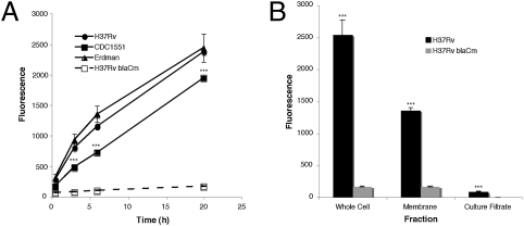
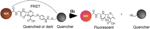
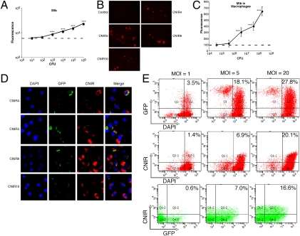
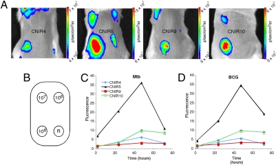
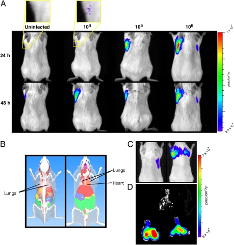
The slow growth rate and genetic intractability of tubercle bacilli has hindered progress toward understanding tuberculosis, one of the most frequent causes of death worldwide. We overcame this roadblock through development of near-infrared (NIR) fluorogenic substrates for beta-lactamase, an enzyme expressed by tubercle bacilli, but not by their eukaryotic hosts, to allow real-time imaging of pulmonary infections and rapid quantification of bacteria in living animals by a strategy called reporter enzyme fluorescence (REF). This strategy has a detection limit of 6 +/- 2 x 10(2) colony-forming units (CFU) of bacteria with the NIR substrate CNIR5 in only 24 h of incubation in vitro, and as few as 10(4) CFU in the lungs of live mice. REF can also be used to differentiate infected from uninfected macrophages by using confocal microscopy and fluorescence activated cell sorting. Mycobacterium tuberculosis and the bacillus Calmette-Guérin can be tracked directly in the lungs of living mice without sacrificing the animals. Therapeutic efficacy can also be evaluated through loss of REF signal within 24 h posttreatment by using in vitro whole-bacteria assays directly in living mice. We expect that rapid quantification of bacteria within tissues of a living host and in the laboratory is potentially transformative for tuberculosis virulence studies, evaluation of therapeutics, and efficacy of vaccine candidates. This is a unique use of an endogenous bacterial enzyme probe to detect and image tubercle bacilli that demonstrates REF is likely to be useful for the study of many bacterial infections.


Conflict of interest statement

The authors declare no conflict of interest.

Figures

Fig. 1.
Fig. 1.
Mycobacterium tuberculosis (Mtb) strains produce a membrane-localized β-lactamase. (A) The three common laboratory Mtb strains H37Rv, CDC1551, and Erdman all produce similar levels of β-lactamase as measured by the change in fluorescence of Fluorocillin Green (excitation: 485 nm; emission: 530 nm) in the presence of 107 bacteria. In the case of CDC1551, the signal produced is significantly lower (***P < 0.001) than Erdman and H37Rv, yet it is at least 10-fold higher than background. The H37Rv blaCm strain with a mutation in the blaC gene displays negligible β-lactamase activity. (B) Membrane fractions of Mtb are responsible for the majority of the β-lactamase activity found in whole-cell fractions, suggesting that BlaC is primarily membrane-localized in Mtb. β-Lactamase levels in culture filtrates are significantly higher than background (P < 0.001), indicating that BlaC is secreted at low levels.
Fig. 2.
Fig. 2.
Reporter enzyme fluorescence detection of M. tuberculosis with near-infrared (CNIR) fluorogenic substrates. The intact CNIR substrates do not display fluorescence due to the close proximity of the fluorescent dye (Cy5 or Cy5.5) and the quencher through fluorescence resonance energy transfer (FRET), but become fluorescent after hydrolysis by β-lactamase, generating the fluorescent product whose emission can be measured using a fluorometer. The quencher groups used are QSY21 or derivatives and the CNIR dyes are Cy5 (CNIR4) and Cy5.5 (CNIR5, CNIR9, and CNIR10). NIR, near-infrared.
Fig. 3.
Fig. 3.
Fluorogenic compounds allow detection of M. tuberculosis (Mtb) in vitro and direct detection of infected cells by confocal microscopy and fluorescence activated cell sorting (FACS). (A) Correlation of fluorescent signal and number of tubercle bacilli in culture as determined by colony-forming units (CFU) in the presence of CNIR5 for 24 h. (B) Fluorescence confocal microscopy of Mtb coincubated with the CNIR substrates for 24 h demonstrates fluorescence incorporation into the bacteria. (C) Correlation of fluorescent signal and number of tubercle bacilli present within J774A.1 murine macrophages after coincubating with CNIR5 for 24 h. (D) Fluorescence confocal microscopy of J774A.1 murine macrophages infected with GFP (green) expressing Mtb and coincubated with CNIR substrates (red) for 24 h. Fixed cells were stained with DAPI (blue) to visualize nuclei. Infected cells can be identified by the presence of signal from CNIR substrates whereas uninfected cells do not incorporate CNIR signal. (E) FACS allows separation of infected J774A.1 murine macrophages labeled with CNIR5 in a manner that correlates well with the multiplicity of infection (MOI) bacteria per cell. Separation can be accomplished with GFP, GFP-expressing Mtb, or CNIR or by using both fluorescent labels. ***P < 0.001: significantly different from fluorescence of medium alone (horizontal dashed lines in A and C) calculated by ANOVA with the Bonferroni posttest.
Fig. 4.
Fig. 4.
Fluorogenic probes allow detection of Mycobacterium tuberculosis (Mtb) and bacillus Calmette–Guérin with similar sensitivity after s.c. infection. (A) Whole-body imaging of mice s.c. infected with Mtb and administered the indicated CNIR substrate. Images were taken 48 h postinfection and administration of the substrate. (B) Inoculation sites, number of bacteria present, and region of interest used for each animal. The position of the reference measurement is indicated (R). (C) Comparison of the signal as a function of time postinjection of each CNIR substrate for 108 Mtb or (D) bacillus Calmette–Guérin.
Fig. 5.
Fig. 5.
Fluorogenic probes allow detection of bacillus Calmette–Guérin and Mycobacterium tuberculosis (Mtb) after pulmonary infection of mice. (A) Signal levels from mice infected with bacillus Calmette–Guérin by the pulmonary route of inoculation that were administered CNIR5 immediately after infection or uninfected mice that were also administered CNIR5. The yellow boxes within the first two mouse image panels indicate the regions that were magnified fourfold to produce the images above the mouse image panels. (B) Depiction of dorsal and ventral views of a mouse with organs to provide the anatomical context for the observed signal. (C) Whole-body images using transillumination after pulmonary infection with Mtb in mice in the same manner as in A. The mouse on the left is uninfected but was administered the CNIR5 substrate whereas the mouse on the right is infected with Mtb by aerosol and was administered CNIR5 immediately after infection. Images were collected at 48 h postinfection with administration of CNIR5 at the same time as infection. (D) Lungs were harvested from infected (lower two sets of lungs) and uninfected (top lungs) animals immediately after live animal images were collected. Lungs were harvested postmortem from the same animals that were first imaged alive (C), and then the lungs themselves were imaged (D). The CFU means and SD of four mice for each dose were (A) 1.3 ± 0.6 × 10⁴, 1.1 ± 0.02 × 10⁵, and 1.0 ± 0.2 × 10⁶ and (C and D) 1.0 ± 0.6 × 10⁶.

References

    1. Dye C, et al. Measuring tuberculosis burden, trends, and the impact of control programmes. Lancet Infect Dis. 2008;8:233–243. - PubMed
    1. Kim DH, et al. Treatment outcomes and long-term survival in patients with extensively drug-resistant tuberculosis. Am J Respir Crit Care Med. 2008;178:1075–1082. - PubMed
    1. Dorman SE, Chaisson RE. From magic bullets back to the magic mountain: The rise of extensively drug-resistant tuberculosis. Nat Med. 2007;13:295–298. - PubMed
    1. Heuts F, Carow B, Wigzell H, Rottenberg ME. Use of non-invasive bioluminescent imaging to assess mycobacterial dissemination in mice, treatment with bactericidal drugs and protective immunity. Microbes Infect. 2009;11:1114–1121. - PubMed
    1. Hickey MJ, et al. Luciferase in vivo expression technology: Use of recombinant mycobacterial reporter strains to evaluate antimycobacterial activity in mice. Antimicrob Agents Chemother. 1996;40:400–407. - PMC - PubMed

Publication types

MeSH terms