

Desulfovibrio magneticus RS-1 contains an iron- and phosphorus-rich organelle distinct from its bullet-shaped magnetosomes
- PMID: 20566879
- PMCID: PMC2901430
- DOI: 10.1073/pnas.1001290107
Desulfovibrio magneticus RS-1 contains an iron- and phosphorus-rich organelle distinct from its bullet-shaped magnetosomes
Abstract
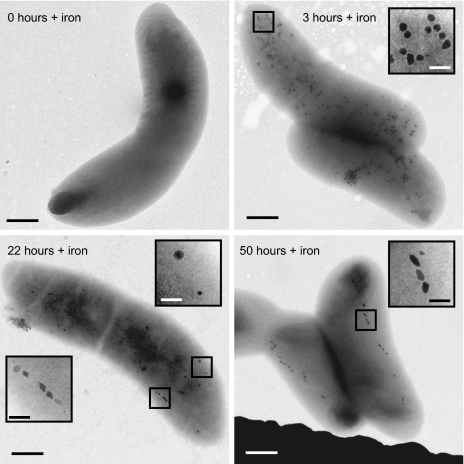
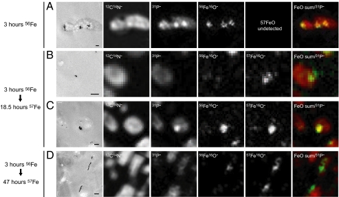
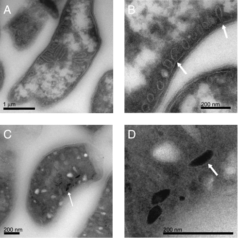
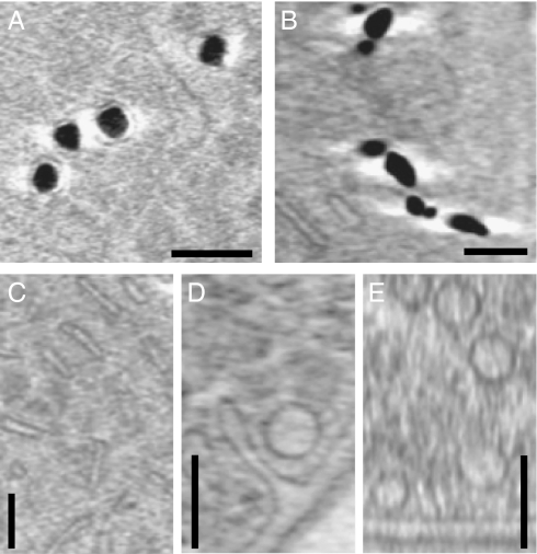
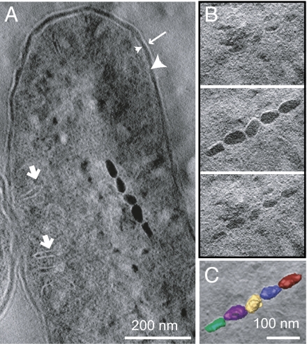
Intracellular magnetite crystal formation by magnetotactic bacteria has emerged as a powerful model for investigating the cellular and molecular mechanisms of biomineralization, a process common to all branches of life. Although magnetotactic bacteria are phylogenetically diverse and their crystals morphologically diverse, studies to date have focused on a few, closely related species with similar crystal habits. Here, we investigate the process of magnetite biomineralization in Desulfovibrio magneticus sp. RS-1, the only reported species of cultured magnetotactic bacteria that is outside of the alpha-Proteobacteria and that forms bullet-shaped crystals. Using a variety of high-resolution imaging and analytical tools, we show that RS-1 cells form amorphous, noncrystalline granules containing iron and phosphorus before forming magnetite crystals. Using NanoSIMS (dynamic secondary ion mass spectroscopy), we show that the iron-phosphorus granules and the magnetite crystals are likely formed through separate cellular processes. Analysis of the cellular ultrastructure of RS-1 using cryo-ultramicrotomy, cryo-electron tomography, and tomography of ultrathin sections reveals that the magnetite crystals are not surrounded by membranes but that the iron-phosphorus granules are surrounded by membranous compartments. The varied cellular paths for the formation of these two minerals lead us to suggest that the iron-phosphorus granules constitute a distinct bacterial organelle.
Conflict of interest statement
The authors declare no conflict of interest.
Figures
Similar articles
-
Genome Editing Method for the Anaerobic Magnetotactic Bacterium Desulfovibrio magneticus RS-1.Appl Environ Microbiol. 2018 Oct 30;84(22):e01724-18. doi: 10.1128/AEM.01724-18. Print 2018 Nov 15. Appl Environ Microbiol. 2018. PMID: 30194101 Free PMC article.
-
Localized iron accumulation precedes nucleation and growth of magnetite crystals in magnetotactic bacteria.Sci Rep. 2017 Aug 15;7(1):8291. doi: 10.1038/s41598-017-08994-9. Sci Rep. 2017. PMID: 28811607 Free PMC article.
-
Proteomic analysis of irregular, bullet-shaped magnetosomes in the sulphate-reducing magnetotactic bacterium Desulfovibrio magneticus RS-1.Proteomics. 2009 Jun;9(12):3341-52. doi: 10.1002/pmic.200800881. Proteomics. 2009. PMID: 19579222
-
The bacterial magnetosome: a unique prokaryotic organelle.J Mol Microbiol Biotechnol. 2013;23(1-2):63-80. doi: 10.1159/000346543. Epub 2013 Apr 18. J Mol Microbiol Biotechnol. 2013. PMID: 23615196 Review.
-
Magnetic microbes: Bacterial magnetite biomineralization.Semin Cell Dev Biol. 2015 Oct;46:36-43. doi: 10.1016/j.semcdb.2015.09.003. Epub 2015 Sep 14. Semin Cell Dev Biol. 2015. PMID: 26382301 Review.
Cited by
-
Magnetite Crystal Orientation in Magnetosome Chains.Adv Funct Mater. 2014 Jul;24(25):3926-3932. doi: 10.1002/adfm.201303737. Epub 2014 Mar 10. Adv Funct Mater. 2014. PMID: 25866495 Free PMC article.
-
Clostridioides difficile ferrosome organelles combat nutritional immunity.Nature. 2023 Nov;623(7989):1009-1016. doi: 10.1038/s41586-023-06719-9. Epub 2023 Nov 15. Nature. 2023. PMID: 37968387 Free PMC article.
-
Phosphorus chemistry and bacterial community composition interact in brackish sediments receiving agricultural discharges.PLoS One. 2011;6(6):e21555. doi: 10.1371/journal.pone.0021555. Epub 2011 Jun 29. PLoS One. 2011. PMID: 21747910 Free PMC article.
-
Bacterial Metallostasis: Metal Sensing, Metalloproteome Remodeling, and Metal Trafficking.Chem Rev. 2024 Dec 25;124(24):13574-13659. doi: 10.1021/acs.chemrev.4c00264. Epub 2024 Dec 10. Chem Rev. 2024. PMID: 39658019 Free PMC article. Review.
-
Calprotectin elicits aberrant iron starvation responses in Pseudomonas aeruginosa under anaerobic conditions.J Bacteriol. 2025 Apr 17;207(4):e0002925. doi: 10.1128/jb.00029-25. Epub 2025 Mar 26. J Bacteriol. 2025. PMID: 40135923 Free PMC article.
References
-
- Bazylinski DA, Frankel RB. Magnetosome formation in prokaryotes. Nat Rev Microbiol. 2004;2:217–230. - PubMed
-
- Crookes-Goodson WJ, Slocik JM, Naik RR. Bio-directed synthesis and assembly of nanomaterials. Chem Soc Rev. 2008;37:2403–2412. - PubMed
-
- Komeili A. Molecular mechanisms of magnetosome formation. Annu Rev Biochem. 2007;76:351–366. - PubMed
Publication types
MeSH terms
Substances
Grants and funding
LinkOut - more resources
Full Text Sources
Medical
Molecular Biology Databases
