. 2010 Jul 6;107(27):12216-21.
doi: 10.1073/pnas.1004143107. Epub 2010 Jun 21.

PDGF-CC blockade inhibits pathological angiogenesis by acting on multiple cellular and molecular targets

Affiliations

PDGF-CC blockade inhibits pathological angiogenesis by acting on multiple cellular and molecular targets

Xu Hou et al. Proc Natl Acad Sci U S A. .

Abstract

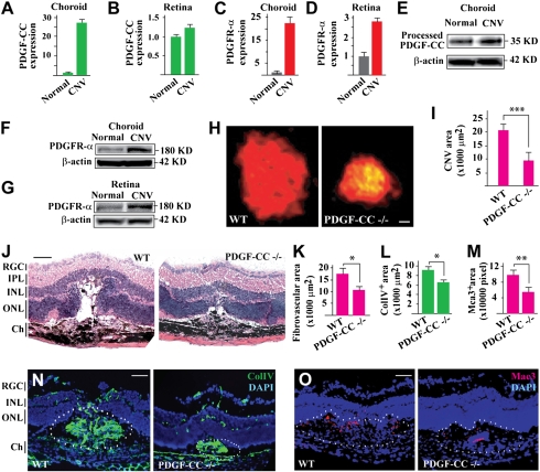
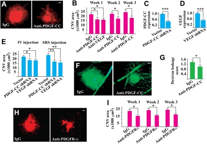
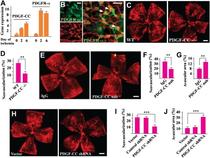
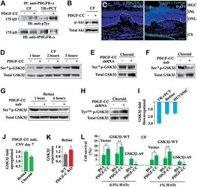
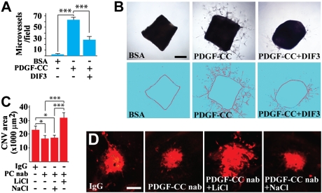
The importance of identifying VEGF-independent pathways in pathological angiogenesis is increasingly recognized as a result of the emerging drug resistance to anti-VEGF therapies. PDGF-CC is the third member of the PDGF family discovered after more than two decades of studies on PDGF-AA and PDGF-BB. The biological function of PDGF-CC and the underlying cellular and molecular mechanisms remain largely unexplored. Here, using different animal models, we report that PDGF-CC inhibition by neutralizing antibody, shRNA, or genetic deletion suppressed both choroidal and retinal neovascularization. Importantly, we revealed that PDGF-CC targeting acted not only on multiple cell types important for pathological angiogenesis, such as vascular mural and endothelial cells, macrophages, choroidal fibroblasts and retinal pigment epithelial cells, but also on the expression of other important angiogenic genes, such as PDGF-BB and PDGF receptors. At a molecular level, we found that PDGF-CC regulated glycogen synthase kinase (GSK)-3beta phosphorylation and expression both in vitro and in vivo. Activation of GSK3beta impaired PDGF-CC-induced angiogenesis, and inhibition of GSK3beta abolished the antiangiogenic effect of PDGF-CC blockade. Thus, we identified PDGF-CC as an important candidate target gene for antiangiogenic therapy, and PDGF-CC inhibition may be of therapeutic value in treating neovascular diseases.


Conflict of interest statement

The authors declare no conflict of interest.

Figures

Fig. 1.
Fig. 1.
Up-regulated PDGF-CC expression in CNV and reduced CNV formation in PDGF-CC–deficient mice. (AD) Real-time PCR showed up-regulated expression of PDGF-CC and PDGFR-α in choroids and retinas 7 d after laser-induced CNV. Arbitrary unit was used for gene expression with the normal controls set to 1. (EG) Western blot confirmed the up-regulated expression of PDGF-CC and PDGFR-α in CNV. (H and I) PDGF-CC deficiency reduced CNV area to approximately 47% of control 7 d after laser treatment as measured by IB4 staining. (J and K) Histological analysis showed less edema formation in the CNV lesions in PDGF-CC–deficient mice as indicated by the reduced empty space around the CNVs. PDGF-CC deficiency also reduced fibrovascular formation. (L and N) PDGF-CC deficiency decreased ColIV+ (vascular marker) areas within the CNV lesions. (M and O) PDGF-CC deficiency decreased Mac3+ (macrophage marker) areas following CNV induction. Blue color in N and O indicates nuclei stained by DAPI. IPL, inner plexiform layer; INL/ONL, inner/outer nuclear layer; Ch, choroid. (Scale bars: 250 μm in H; 50 μm in J, N, and O.) *P < 0.05, **P < 0.01, ***P < 0.001.
Fig. 2.
Fig. 2.
PDGF-CC targeting inhibited CNV formation and blood vessel leakage. (A and B) PDGF-CC–neutralizing antibody treatment reduced CNV areas at different time points with a comparable effect of a VEGF-neutralizing antibody. (CE) Treatment with PDGF-CC shRNA, which reduced PDGF-CC expression level to approximately 40% of normal level (C), reduced CNV areas 7 d after laser treatment when injected into vitreous (IV) or subretinal space (SRS) (E). The effect of the PDGF-CC shRNA was comparable to that of a VEGF shRNA (E), which reduced VEGF expression level to approximately 40% of normal level (D). (F and G) Fluorescein angiography showed reduced blood vessel leakage in the PDGF-CC–neutralizing antibody–treated CNVs 1 wk after laser treatment. (H and I) Intravitreal injection of PDGFR-α–neutralizing antibody decreased CNV areas at different time points after laser treatment. The inhibitory effect of PDGFR-α–neutralizing antibody was similar to that of PDGF-CC–neutralizing antibody. (Scale bar: 50 μm in A, F, and H.) *P < 0.05, **P < 0.01, ***P < 0.001.
Fig. 3.
Fig. 3.
PDGF-CC deficiency and blockades inhibited ischemia-induced retinal neovascularization. (A) Real-time PCR showed up-regulated expression of PDGF-CC and PDGFR-α during retinal ischemia. Arbitrary unit was used for gene expression with the d 0 control set to 1. (B) Immunofluorescence staining showed PDGFR-α expression by the neovessels in the retina, with a higher expression level in the neovascular tufts. (C and D) PDGF-CC deficiency reduced retinal neovascularization 5 d after retinal ischemia. (EG) PDGF-CC nAb intravitreal injection inhibited retinal neovascularization (E and F), leading to greater avascular areas in the retinas (E and G). (HJ) Intravitreal injection of PDGF-CC shRNA decreased retinal neovascularization (H and I), leading to greater avascular areas in the retinas (H and J). (Scale bars: 100 μm in B; 500 μm in C, E, and H.) ** P < 0.01, *** P < 0.001.
Fig. 4.
Fig. 4.
PDGF-CC activated PDGFR-α, Akt, regulated GSK3β phosphorylation/expression, and GSK3β Ser9 phosphorylation was required for the survival effect of PDGF-CC. (A and B) IP followed by IB showed that PDGF-CC protein stimulation activated PDGFR-α in mCFs and TR-rPCTs (A) and activated Akt in mCFs (B). (C) Immunofluorescence staining detected p-PDGFR-α (green) within the CNV area (lined). Blue color indicates nuclei stained by DAPI. IPL, inner plexiform layer; ONL, outer nuclear layer; Ch, choroid. (Scale bar: 50 μm.) (D) PDGF-CC protein increased Ser9 GSK3β phosphorylation in mCFs in a time-dependent manner. (E and F) PDGF-CC shRNA (E) and nAb (F) treatment reduced GSK3β Ser9 phosphorylation in the choroids in vivo. (G) PDGF-CC–neutralizing antibody treatment reduced GSK3β Ser9 phosphorylation in the mouse retina in vivo. (H) PDGF-CC shRNA treatment increased GSK3β Tyr216 phosphorylation in mouse choroids in vivo. (I) PDGF-CC protein treatment inhibited GSK3β expression in cultured vascular cells in vitro (e.g., TR-rPCT, HUVSMC) and in the retina in vivo as measured by real-time PCR. (J) PDGF-CC–neutralizing antibody treatment increased GSK3β expression in choroids and retinas with CNV as measured by real-time PCR. (K) Real-time PCR showed that GSK3β expression level was approximately 35% higher in the PDGF-CC–deficient retinas than that of normal control. (L) PDGF-CC protein treatment protected CFs from H2O2-induced cell death in the WT GSK3β– and vector-transfected cells. The survival effect of PDGF-CC diminished in the GSK3β-A9–transfected cells. *P < 0.05, **P < 0.01.
Fig. 5.
Fig. 5.
Involvement of GSK3β in PDGF-CC–induced angiogenesis and in the antiangiogenic effect of PDGF-CC targeting. (A and B) PDGF-CC protein induced vascular cell proliferation, migration, and microvessel formation in the aortic ring assay (B, Middle). Cotreatment of the aortic rings with the GSK3β activator DIF3 impaired PDGF-CC–induced microvessel formation (B, Right). (Scale bar: 500 μm.) (C and D) Intravitreal injection of PDGF-CC nAb (PC nAb) inhibited CNV formation. Coinjection of the GSK3β inhibitor LiCl abolished the PDGF-CC–neutralizing antibody induced reduction of CNV. NaCl was used as a control and had no effect. (Scale bars: 100 μm.) *P < 0.05, ***P < 0.001.

Similar articles

Cited by

References

    1. Ferrara N. Pathways mediating VEGF-independent tumor angiogenesis. Cytokine Growth Factor Rev. 2010;21:21–26. - PubMed
    1. Benjamin LE, Hemo I, Keshet E. A plasticity window for blood vessel remodelling is defined by pericyte coverage of the preformed endothelial network and is regulated by PDGF-B and VEGF. Development. 1998;125:1591–1598. - PubMed
    1. Crawford Y, Ferrara N. Tumor and stromal pathways mediating refractoriness/resistance to anti-angiogenic therapies. Trends Pharmacol Sci. 2009;30:624–630. - PubMed
    1. Maharaj AS, et al. VEGF and TGF-beta are required for the maintenance of the choroid plexus and ependyma. J Exp Med. 2008;205:491–501. - PMC - PubMed
    1. Li X, et al. PDGF-C is a new protease-activated ligand for the PDGF alpha-receptor. Nat Cell Biol. 2000;2:302–309. - PubMed

Publication types

MeSH terms