

Quantitative proteomics reveals myosin and actin as promising saliva biomarkers for distinguishing pre-malignant and malignant oral lesions
- PMID: 20567502
- PMCID: PMC2887353
- DOI: 10.1371/journal.pone.0011148
Quantitative proteomics reveals myosin and actin as promising saliva biomarkers for distinguishing pre-malignant and malignant oral lesions
Abstract
Background: Oral cancer survival rates increase significantly when it is detected and treated early. Unfortunately, clinicians now lack tests which easily and reliably distinguish pre-malignant oral lesions from those already transitioned to malignancy. A test for proteins, ones found in non-invasively-collected whole saliva and whose abundances distinguish these lesion types, would meet this critical need.
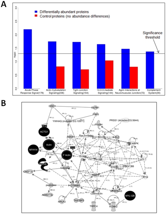
Methodology/principal findings: To discover such proteins, in a first-of-its-kind study we used advanced mass spectrometry-based quantitative proteomics analysis of the pooled soluble fraction of whole saliva from four subjects with pre-malignant lesions and four with malignant lesions. We prioritized candidate biomarkers via bioinformatics and validated selected proteins by western blotting. Bioinformatic analysis of differentially abundant proteins and initial western blotting revealed increased abundance of myosin and actin in patients with malignant lesions. We validated those results by additional western blotting of individual whole saliva samples from twelve other subjects with pre-malignant oral lesions and twelve with malignant oral lesions. Sensitivity/specificity values for distinguishing between different lesion types were 100%/75% (p = 0.002) for actin, and 67%/83% (p<0.00001) for myosin in soluble saliva. Exfoliated epithelial cells from subjects' saliva also showed increased myosin and actin abundance in those with malignant lesions, linking our observations in soluble saliva to abundance differences between pre-malignant and malignant cells.
Conclusions/significance: Salivary actin and myosin abundances distinguish oral lesion types with sensitivity and specificity rivaling other non-invasive oral cancer tests. Our findings provide a promising starting point for the development of non-invasive and inexpensive salivary tests to reliably detect oral cancer early.
Conflict of interest statement
Figures


References
-
- Rhodus NL. Oral Cancer: Early Detection and Prevention. Inside Dentistry. 2007;3:1–8.
-
- Bethesda, MD: National Cancer Institute; 1993. SEER Cancer Statistics Review 1975-2006.
-
- Hofman LF. Human saliva as a diagnostic specimen. J Nutr. 2001;131:1621S–1625S. - PubMed
-
- Wong DT. Towards a simple, saliva-based test for the detection of oral cancer ‘oral fluid (saliva), which is the mirror of the body, is a perfect medium to be explored for health and disease surveillance’. Expert Rev Mol Diagn. 2006;6:267–272. - PubMed
Publication types
MeSH terms
Substances
Grants and funding
LinkOut - more resources
Full Text Sources
Medical
