

Investigation of gold-coated bare fiber probe for in situ intra-vitreous coherence domain optical imaging and sensing
- PMID: 20567605
- PMCID: PMC2887671
- DOI: 10.1007/s00340-010-3910-4
Investigation of gold-coated bare fiber probe for in situ intra-vitreous coherence domain optical imaging and sensing
Abstract
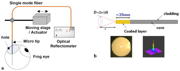
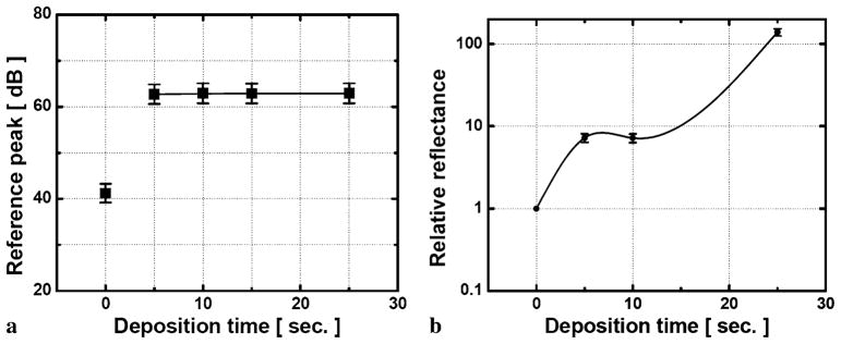
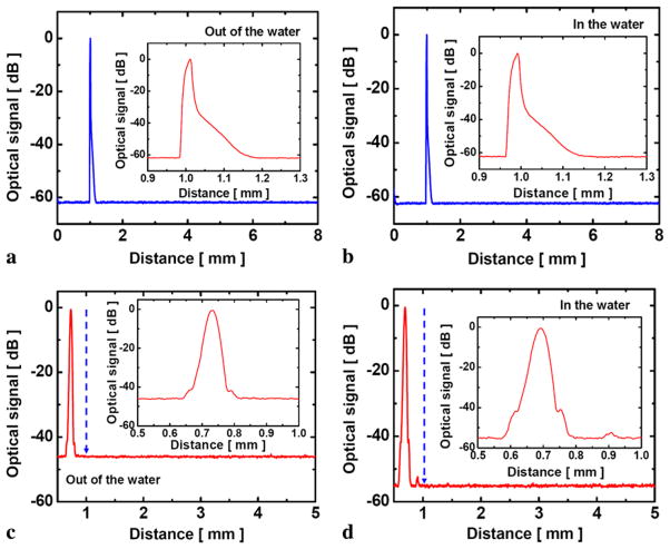
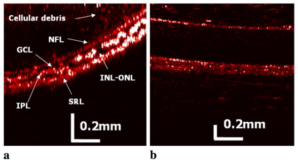
We have investigated the usage of gold-plated bare fiber probes for in situ imaging of retinal layers and surrounding ocular tissues using time-domain common-path optical coherence tomography. The fabricated intra-vitreous gold-plated micro-fiber probe can be fully integrated with surgical tools working in close proximity to the tissue to provide subsurface images having a self-contained reference plane independent to the Fresnel reflection between the distal end of the probe and the following medium for achieving reference in typical common-path optical coherence tomography. We have fully characterized the probe in an aqueous medium equivalent to the vitreous humor in the eye and were able to differentiate various functional retinal tissue layers whose thickness is larger than the system's resolution.
Figures
Similar articles
-
Sapphire ball lensed fiber probe for common-path optical coherence tomography in ocular imaging and sensing.Proc SPIE Int Soc Opt Eng. 2013 Mar 26;8567:10.1117/12.2005099. doi: 10.1117/12.2005099. Proc SPIE Int Soc Opt Eng. 2013. PMID: 24392202 Free PMC article.
-
In-line optical fiber metallic mirror reflector for monolithic common path optical coherence tomography probes.Lasers Surg Med. 2018 Mar;50(3):230-235. doi: 10.1002/lsm.22756. Epub 2017 Nov 6. Lasers Surg Med. 2018. PMID: 29105794
-
Common-path optical coherence tomography with side-viewing bare fiber probe for endoscopic optical coherence tomography.Rev Sci Instrum. 2007 Nov;78(11):113102. doi: 10.1063/1.2804112. Rev Sci Instrum. 2007. PMID: 18052460
-
Ultra high-resolution anterior segment optical coherence tomography in the diagnosis and management of ocular surface squamous neoplasia.Ocul Surf. 2014 Jan;12(1):46-58. doi: 10.1016/j.jtos.2013.11.001. Epub 2013 Nov 9. Ocul Surf. 2014. PMID: 24439046 Free PMC article. Review.
-
[Optical coherence tomography, an important new tool in the investigation of the retina].Ned Tijdschr Geneeskd. 2005 Aug 20;149(34):1884-91. Ned Tijdschr Geneeskd. 2005. PMID: 16136741 Review. Dutch.
Cited by
-
Etching-enabled extreme miniaturization of graded-index fiber-based optical coherence tomography probes.J Biomed Opt. 2019 Nov;25(3):1-5. doi: 10.1117/1.JBO.25.3.032006. J Biomed Opt. 2019. PMID: 31707773 Free PMC article.
-
Common-Path Optical Coherence Tomography for Biomedical Imaging and Sensing.J Opt Soc Korea. 2010 Mar;14(1):1-13. doi: 10.3807/JOSK.2010.14.1.001. J Opt Soc Korea. 2010. PMID: 20657808 Free PMC article.
References
-
- Ikeda F, Iida T, Kishi S. Ophthalmology. 2008;115:718. - PubMed
-
- Sayanagi K, Sharma S, Kaiser PK. Ophthalmic Surg Lasers Imaging. 2009;40:195. - PubMed
-
- Wong HT, Lim MC, Sakata LM, Aung HT, Amerasinghe N, Friedman DS, Aung T. Arch Ophthalmol. 2009;127:256. - PubMed
-
- Kim SJ, Bressler NM. Curr Opin Ophthalmol. 2009;20:46. - PubMed
-
- Feltgen N, Junker B, Agostini H, Hansen L. Ophthalmology. 2007;114:716. - PubMed
Grants and funding
LinkOut - more resources
Full Text Sources
Other Literature Sources